
Page 89 - Read Online
P. 89
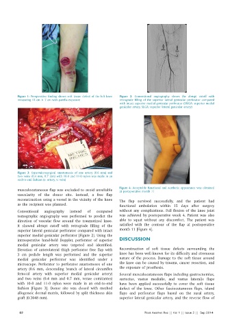
Figure 1: Preoperative finding shows soft tissue defect of the left knee   Figure 2:  Conventional angiography shows the  abrupt cutoff with
          measuring 12 cm × 7 cm with patella exposure        retrograde filling of the superior lateral genicular perforator  compared
                                                              with intact superior medial genicular perforator (SMGA: superior medial
                                                              genicular artery, SLGA: superior lateral genicular artery)


          Figure  3:  Supermicrosurgical anastomosis of one artery (0.6 mm) and
          two veins (0.4 mm, 0.7 mm) with 10‑0 and 11‑0 nylon was made in an
          end‑to‑end fashion (a: artery, v: vein)
                                                              Figure 4:  Acceptable functional and aesthetic  appearance was obtained
          musculocutaneous flap was excluded to avoid unreliable   at postoperative month 11
          vascularity  of  the  donor  site.  Instead,  a  free  flap
          reconstruction using a vessel in the vicinity of the knee   The  flap survived  successfully,  and the  patient  had
          as the recipient was planned.                       functional ambulation within 15  days after surgery

          Conventional  angiography  instead  of  computed    without any complications. Full flexion of the knee joint
          tomographic angiography was performed to predict the   was achieved  by  postoperative  week 4.  Patient  was  also
          direction of vascular flow around the traumatized  knee.   able to squat without any discomfort. The patient was
          It showed abrupt cutoff with retrograde filling of the   satisfied  with the contour of the flap at postoperative
          superior lateral genicular perforator compared with intact   month 11 [Figure 4].
          superior medial genicular perforator [Figure 2]. Using the
          intraoperative hand‑held  Doppler, perforator  of superior   DISCUSSION
          medial  genicular artery  was targeted  and identified.
          Elevation  of anterolateral thigh  perforator free  flap with   Reconstruction of soft tissue defects surrounding the
          3  cm pedicle length was performed and the superior   knee has been well-known for its difficulty and strenuous
          medial genicular perforator was identified under a   nature of the process. Damage to the soft tissue around
          microscope. Perforator to perforator anastomoses of one   the knee can be caused by trauma, cancer resection, and
          artery  (0.6  mm,  descending  branch of  lateral  circumflex   the exposure of prosthesis.
          femoral artery with superior medial genicular artery)   Several musculocutaneous flaps including gastrocnemius,
          and two veins  (0.4  mm  and 0.7  mm,  venae  comitantes)   sartorius, vastus medialis, and vastus lateralis flaps
          with 10-0 and 11-0 nylon were made in an end-to-end   have been applied successfully to cover the soft tissue
          fashion  [Figure  3]. Donor  site was closed  with meshed   defect of the knee. Other fasciocutaneous flaps, island
          allogeneic dermal matrix, followed by split thickness skin   flaps and perforator flaps based on the sural artery,
          graft (0.3048 mm).                                  superior lateral genicular artery, and the reverse flow of


            82                                                             Plast Aesthet Res || Vol 1 || Issue 2 ||  Sep 2014