
Page 77 - Read Online
P. 77
the incisions are completed, and the flap islanded. The
inner curvilinear edge of the keystone flap is advanced
medially for coverage of the defect. An advancement
of 3 cm can be obtained; further advancement would
require skeletonization of the perforators. The defect
is narrowed by closing either ends in a V‑Y fashion.
This redistributes tension on the inset also. Interrupted
simple sutures are placed. We do not require elaborate
suturing (in the HEMMING pattern) as is done in the
classical keystone‑design. Primary closure of the
[2]
secondary defect can be achieved especially in the upper
leg. In the case of the lower leg, closure requires a skin
graft. Two clinical examples are illustrated.
RESULTS a b
Figure 1: (a) When the defect is on the upper leg, the flap is designed
on the medial calf region, and is advanced medially. (b) when the defect
The patient with squamous cell carcinoma is on the lower leg, the flap is designed on the lateral side, and is
A 50‑year‑old woman underwent wide local excision advanced medially
of squamous cell carcinoma over the pretibial region of
her left leg [Figure 2]. A 20 cm × 9 cm keystone‑design
perforator‑based flap was marked over the medial calf
after identifying three perforators with Doppler. These
were found to arise from the medial sural artery on a b
exploration. The flap was islanded on these perforators
and advanced medially to cover the tibia. Part of the
primary defect medial to the exposed bone was skin‑
grafted. The secondary defect was closed primarily. c d
Healing was uneventful, and the patient is asymptomatic,
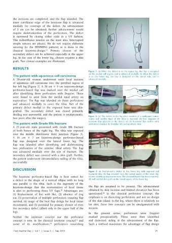
two years after the surgery. Figure 2: (a) The defect in the leg after excision of a malignant tumor.
Upper and middle third of the tibia is exposed. (b) line diagram of
keystone flap adjacent to the defect. (c) the keystone flap outline with
The patient with Grade IIIb fracture perforators marked by Doppler. (d) postoperative view
A 21‑year‑old male presented with Grade IIIb fracture
of both bones of the right leg. The tibia was exposed
over the middle third‑lower third junction [Figure 3].
A 16 cm × 7 cm keystone‑design perforator‑based
flap was designed over the lateral lower leg. The
flap was islanded after identifying and skeletonizing
two perforators of the anterior tibial artery. The flap a b
was advanced medially over the site of fracture. The
secondary defect was covered with a skin graft. Further,
the patient underwent intramedullary nailing of the tibia,
successfully.
c d
DISCUSSION
Figure 3: (a) Posttraumatic defect in the lower leg with exposed and
fractured tibia, (b) flap elevated from the lateral aspect of the lower leg.
The keystone perforator‑based flap is best suited for (c) two months postoperative view. The external fixator has been removed.
a defect in the shape of a vertical ellipse with its long (d) well settled skin graft on the lateral aspect of the lower leg
axis parallel to the tibia. Such is the ingenuity of the
keystone‑design that the reorientation of local tissue the flap are assumed to be present. The advancement
is akin to performing three V‑Y flaps. Advantages are: obtained by skin incision and limited elevation has been
[3]
[5]
(1) Replacement of like with like, (2) absence of dog questioned. In the classical perforator concept, the
ear, (3) preservation of multiple perforators ensuring flap emphasis is on dissecting perforators and not on design
survival, (4) usage of the best flap design for local tissue of the skin island. In the leg, where there is relatively no
recruitment, and (5) potential for primary closure of even lax skin, these two concepts can be amalgamated with
the secondary defect (albeit only in the upper half of the success.
leg). In the present series, perforators were Doppler
Neither the keystone concept nor the perforator marked preoperatively. These were then identified
concept is new. In the classical keystone concept and and dissected, aiding in the advancement of the flap.
[1]
some of its modifications, perforators nourishing Such a method maximizes the advantage of flap design
[4]
Plast Aesthet Res || Vol 1 || Issue 2 || Sep 2014 71