
Page 72 - Read Online
P. 72
of which are important in modulating mesenchymal RESULTS
cell recruitment, proliferation, and extracellular matrix
[4]
synthesis during the healing process. Autologous PRP is Twenty‑four patients with 33 nonhealing ulcers of various
a safe, easy, and cost‑effective method with good results etiologies were treated with PRP at weekly intervals for a
in the management of chronic nonhealing ulcers. PRP has maximum of 6 treatments. The mean age of the patients
been a breakthrough in the stimulation and acceleration was 42.5 years (standard deviation [SD] 12.48) [Table 1]. Of
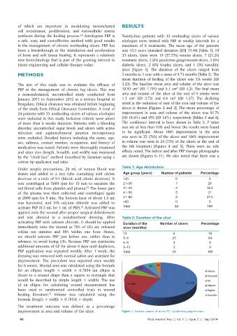
of bone and soft tissue healing. It represents a relatively 33 ulcers, there were 19 (57.75%) venous ulcers, 7 (21.2%)
new biotechnology that is part of the growing interest in traumatic ulcers, 2 (6%) pyoderma gangrenosum ulcers, 2 (6%)
tissue engineering and cellular therapy today. diabetic ulcers, 2 (6%) trophic ulcers, and 1 (3%) vasculitic
ulcer [Figure 1]. The duration of the ulcers ranged from
METHODS 2 months to 1 year with a mean of 4.75 months [Table 2]. The
mean duration of healing of the ulcers was 5.6 weeks (SD
The aim of this study was to evaluate the efficacy of 3.23). The baseline mean area and volume of the ulcer was
2
3
PRP in the management of chronic leg ulcers. This was 10.93 cm (SD 7.791) and 5.1 cm (SD 4.3). The final mean
a nonrandomized, uncontrolled study conducted from area and volume of the ulcer at the end of 6 weeks were
2
3
January 2011 to September 2012 at a tertiary hospital in 1.3 cm (SD 2.72) and 0.4 cm (SD 1.27). The declining
Bengaluru. Ethical clearance was obtained before beginning trend in the reduction of sum of the area and volume of the
of the study from Ethical Clearance Committee. A total of ulcers is shown [Figures 2 and 3]. The mean percentage of
24 patients with 33 nonhealing ulcers of various etiologies improvement in area and volume of the ulcers was 91.7%
were included in this study. Inclusion criteria were ulcers (SD 18.4%) and 95% (SD 14%), respectively [Tables 3 and 4].
of more than 6 weeks duration. Patients with a bleeding The confidence interval is been shown in Table 5. P value
disorder, uncontrolled sugar levels and ulcers with active was set at less than 0.05 and hence the results were found
infection and saphenofemoral junction incompetency to be significant. About 100% improvement in the area
were excluded. Detailed history including the name, age, was seen in 25 (76%) of the ulcers and 100% improvement
sex, address, contact number, occupation, and history of in volume was seen in 24 (73%) of the ulcers at the end of
medication was noted. Patients were thoroughly examined the 6th treatment [Figures 4 and 5]. There were no side
and ulcer size (length, breadth, and width) was measured effects noted. The before and after PRP therapy photographs
by the “clock‑face” method described by Sussman using a are shown [Figures 6–11]. We also noted that there was a
cotton tip applicator and ruler.
Table 1: Age distribution
Under aseptic precautions, 20 mL of venous blood was
drawn and added to a test tube containing acid citrate Age group (years) Number of patients Percentage
dextrose in a ratio of 9:1 (blood: acid citrate dextrose). It <20 0 0
was centrifuged at 5000 rpm for 15 min to separate the 21–30 7 29
[5]
red blood cells from platelet and plasma. The lower part 31–40 3 12.5
of the plasma was then collected and centrifuged again 41–50 5 21
at 2000 rpm for 5 min. The bottom layer of about 1.5 mL 51–60 9 37.5
was harvested, and 10% calcium chloride was added to >60 0 0
activate PRP (0.3 mL for 1 mL of PRP). Activated PRP was Total 24 100
[6]
applied onto the wound after proper surgical debridement
and was dressed in a nonabsorbent dressing. After Table 2: Duration of the ulcer
activating PRP with calcium chloride, it should be applied Duration of the Number of ulcers Percentage
immediately onto the wound as 70% of GFs are released ulcer (months)
within ten minutes and 90% within one hour. Hence, <3 6 18
we should activate PRP just before use, rather than in 3–6 21 64
advance, to avoid losing GFs. Because PRP can synthesize 6–9 3 9
additional amounts of GF for about 8 days until depletion, 9–12 3 9
PRP application was repeated weekly. After 1 week, the Total 33 100
dressing was removed with normal saline and assessed for
improvement. The procedure was repeated once weekly
for 6 weeks. Wound area was calculated using the formula
for an ellipse: length × width × 0.7854 (an ellipse is
closer to a wound shape than a square or rectangle that
would be described by simple length × width). The use
of an ellipse for calculating wound measurement has
been used in randomized controlled trials in wound
healing literature. Volume was calculated using the
[7]
formula (length × width × 0.7854) × depth.
The treatment outcome was defined as a percentage
improvement in area and volume of the ulcer. Figure 1: Various causes of ulcer. PG: pyoderma gangrenosum
66 Plast Aesthet Res || Vol 1 || Issue 2 || Sep 2014