
Page 53 - Read Online
P. 53
a b
Figure 3: Filopodia of endothelial cells induced by nanotopography
[27]
(used with permission)
dentistry through the aid of nanorobotics, nanomaterials,
and biotechnology. Nanorobots have a diameter of
[33]
0.5–3 µm and are made of components sized from 1 nm
to 100 nm. They can be programmed, thus enabling
clinicians to execute accurate procedures at the cellular c
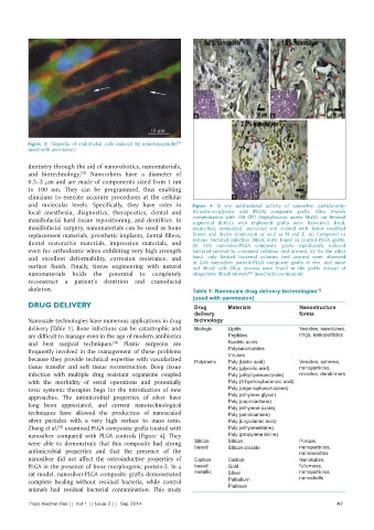
and molecular levels. Specifically, they have roles in Figure 4: In vivo antibacterial activity of nanosilver particle‑poly‑
local anesthesia, diagnostics, therapeutics, dental and DL‑lactic‑co‑glycolic acid (PLGA) composite grafts. After 2‑week
maxillofacial hard tissue repositioning, and dentifrice. In contamination with 108 CFU Staphylococcus aureus Mu50, rat femoral
segmental defects with implanted grafts were harvested, fixed,
maxillofacial surgery, nanomaterials can be used as bone decalcified, embedded, sectioned and stained with Taylor modified
replacement materials, prosthetic implants, dental fillers, Brown and Brenn Gram‑stain as well as H and E. (a) Compared to
dental restorative materials, impression materials, and serious bacterial infection (black dots) found in control PLGA grafts,
(b) 1.0% nanosilver‑PLGA composite grafts significantly reduced
even for orthodontic wires exhibiting very high strength bacterial survival to colonized collation (red arrows). (c) On the other
and excellent deformability, corrosion resistance, and hand, only limited bacterial colonies (red arrows) were observed
surface finish. Finally, tissue engineering with natural in 2.0% nanosilver particle‑PLGA composite grafts in vivo, and more
red blood cells (blue arrows) were found in the grafts instead of
nanomaterials holds the potential to completely phagocytes (black arrows) (used with permission)
[35]
reconstruct a patient’s dentition and craniofacial
skeleton. Table 1: Nanoscale drug delivery technologies
[1]
(used with permission)
DRUG DELIVERY Drug Materials Nanostructure
delivery forms
Nanoscale technologies have numerous applications in drug technology
delivery [Table 1]. Bone infections can be catastrophic and Biologic Lipids Vesicles, nanotubes,
are difficult to manage even in the age of modern antibiotics Peptides rings, nanoparticles
and best surgical techniques. Plastic surgeons are Nucleic acids
[34]
frequently involved in the management of these problems Polysaccharides
because they provide technical expertise with vascularized Polymeric Viruses Vesicles, spheres,
Poly (lactic acid)
tissue transfer and soft tissue reconstruction. Deep tissue Poly (glycolic acid) nanoparticles,
infection with multiple drug resistant organisms coupled Poly (alkylcyanoacrylate) micelles, dendrimers
with the morbidity of serial operations and potentially Poly (3-hydroxybutanoic acid)
toxic systemic therapies begs for the introduction of new Poly (organophosphazene)
approaches. The antimicrobial properties of silver have Poly (ethylene glycol)
long been appreciated, and current nanotechnological Poly (caprolactone)
Poly (ethylene oxide)
techniques have allowed the production of nanoscaled Poly (amidoamine)
silver particles with a very high surface to mass ratio. Poly (L-glutamic acid)
Zheng et al. examined PLGA composite grafts treated with Poly (ethyleneimine)
[35]
nanosilver compared with PLGA controls [Figure 4]. They Poly (propylene imine)
were able to demonstrate that this composite had strong Silicon Silicon Porous,
nanoparticles,
based
antimicrobial properties and that the presence of the Silicon dioxide nanoneedles
nanosilver did not affect the osteoinductive properties of Carbon Carbon Nanotubes,
PLGA in the presence of bone morphogenic protein‑2. In a based Gold fullerness,
rat model, nanosilver‑PLGA composite grafts demonstrated metallic Silver nanoparticles,
complete healing without residual bacteria, while control Palladium nanoshells
animals had residual bacterial contamination. This study Platinum
Plast Aesthet Res || Vol 1 || Issue 2 || Sep 2014 47