
Page 51 - Read Online
P. 51
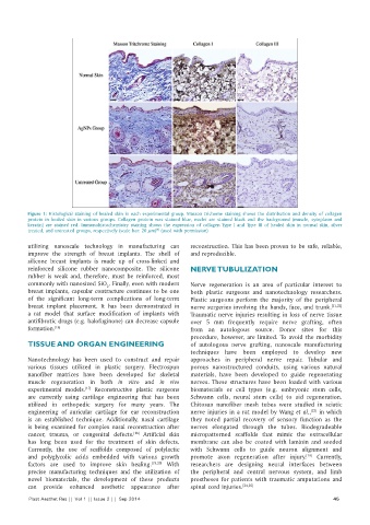
Figure 1:  Histological staining  of healed skin  in  each experimental  group. Masson  trichome  staining  shows  the  distribution  and density  of collagen
          protein  in  healed skin  in  various groups.  Collagen protein  was  stained  blue,  nuclei are  stained  black and the  background (muscle, cytoplasm and
          keratin) are stained red. Immunohistochemistry staining  shows the expression of collagen Type I and Type III of healed skin in normal skin, silver
                                                   [9]
          treated, and untreated groups, respectively (scale bar: 20 µm)  (used with permission)
          utilizing  nanoscale technology in manufacturing can   reconstruction. This has been proven to be safe, reliable,
          improve  the  strength  of breast  implants.  The  shell  of   and reproducible.
          silicone breast implants is made up of cross‑linked and
          reinforced silicone  rubber  nanocomposite. The silicone   NERVE TUBULIZATION
          rubber  is  weak and, therefore,  must  be  reinforced, most
          commonly with nanosized SiO . Finally, even with modern   Nerve regeneration is an area of particular interest to
                                   2
          breast implants, capsular contracture continues to be one   both plastic surgeons and nanotechnology researchers.
          of the  significant  long‑term  complications of long‑term   Plastic surgeons perform the majority of the peripheral
          breast  implant  placement.  It  has  been  demonstrated  in   nerve surgeries involving the hands, face, and trunk. [21,22]
          a rat model that surface modification of implants with   Traumatic nerve injuries resulting in loss of nerve tissue
          antifibrotic drugs (e.g. halofuginone) can decrease capsule   over  5  mm frequently require nerve grafting, often
          formation. [16]                                     from an autologous source. Donor sites for this
                                                              procedure, however, are limited. To avoid the morbidity
          TISSUE AND ORGAN ENGINEERING                        of  autologous  nerve  grafting,  nanoscale  manufacturing
                                                              techniques have been employed to develop new
          Nanotechnology has been used to construct and repair   approaches in peripheral nerve repair. Tubular and
          various tissues  utilized in plastic surgery. Electrospun   porous nanostructured conduits, using various natural
          nanofiber matrices  have been developed for skeletal   materials,  have  been  developed  to  guide  regenerating
          muscle regeneration in both  in  vitro  and  in  vivo   nerves. These structures have been loaded with various
          experimental  models.  Reconstructive plastic surgeons   biomaterials or cell types  (e.g.  embryonic stem cells,
                            [17]
          are  currently  using  cartilage engineering  that  has been   Schwann cells, neural stem cells) to aid regeneration.
          utilized  in  orthopedic surgery  for  many  years.  The   Chitosan nanofiber mesh tubes were studied in sciatic
          engineering  of auricular cartilage for ear reconstruction   nerve injuries in a rat model by Wang et al.,  in which
                                                                                                     [23]
          is  an  established  technique.  Additionally,  nasal  cartilage   they noted partial recovery of sensory function as the
          is being examined for complex nasal reconstruction after   nerves elongated through the tubes. Biodegradeable
          cancer, trauma,  or congenital defects.  Artificial skin   micropatterned scaffolds that mimic the extracellular
                                            [18]
          has  long been  used  for the  treatment  of skin  defects.   membrane  can  also  be  coated  with  laminin  and  seeded
          Currently, the use of scaffolds  composed  of polylactic   with Schwann cells to guide neuron alignment and
          and polyglycolic acids embedded with various growth   promote axon regeneration after injury.  Currently,
                                                                                                    [19]
          factors are  used  to improve  skin  healing. [19,20]   With   researchers are designing neural interfaces between
          precise manufacturing techniques  and the utilization  of   the peripheral and central nervous system, and limb
          novel biomaterials,  the  development of these  products   prostheses for patients with traumatic amputations and
          can provide enhanced aesthetic  appearance after    spinal cord injuries. [24,25]
          Plast Aesthet Res || Vol 1 || Issue 2 ||  Sep 2014                                                45