
Page 52 - Read Online
P. 52
NANOTECHNOLOGY IN BONE had improved load‑bearing limitations. This improved
BIOLOGY AND REPAIR mechanical strength was most likely due to the strong
bonds between nanoparticles and PGLA, as conveyed by
Current developments in bone matrix depend on the the fine ultrastructure of the particles. This enhancement
understanding that the bone microenvironment is made of mechanical strength, through the application of
up of progenitor cells, mineralized ECM scaffold, soluble nanoparticles, is a previously underappreciated finding
chemical signals (such as cytokines), and mechanical in nanomaterials. Finally, this work highlights that the
stimuli. [26] Nanoscale fabrication techniques can three‑dimensional structure of nanoparticles and its
improve each of these components. Scaffolds made of interactions can increase their applications.
nanomaterials provide a geometric porous structure that Hydroxyapatite (HA) is currently used to fill bone defects by
allows osteoblastic differentiation. Such techniques are itself or as a prosthetic coating. While HA has advantages
[27]
conceptually simple, yet were not technically possible over other bioceramics, such as creating strong bonds with
until the development of modern nanoscale fabrication native tissues, it lacks a homogeneous degradation phase.
techniques. Advances in fabrication and manufacturing Given the nanoscale architecture of native bone crystals,
make nanotechnology an exciting and powerful tool in manufacturing HA on a nanoscale would theoretically
the development of bone reconstruction. improve its utility. Poinern et al. investigated the effects
[30]
of thermal and ultrasonic techniques for the development
BONE PROSTHESES of these particles and demonstrated that either technique
can generate particles of similar consistency. Abd El‑Fattah
Nanotechnology can be used to manipulate the surfaces et al. histomorphometrically analyzed the tissue by
[31]
of standard bone replacement implants to maximize tissue growth and scaffold degradation in three groups of rats
ingrowth, while minimizing inflammation. Raimondo et al. with identical bone defects: one filled with mirco‑HA,
[28]
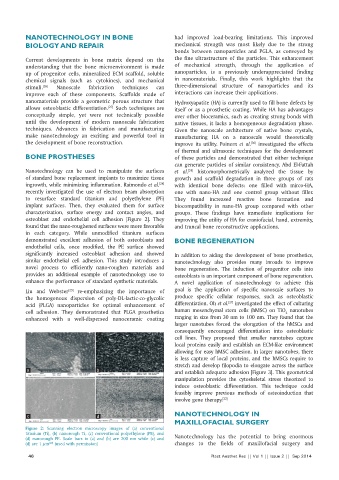
recently investigated the use of electron beam absorption one with nano‑HA and one control group without filler.
to resurface standard titanium and polyethylene (PE) They found increased reactive bone formation and
implant surfaces. Then, they evaluated them for surface biocompatibility in nano‑HA group compared with other
characterization, surface energy and contact angles, and groups. These findings have immediate implications for
osteoblast and endothelial cell adhesion [Figure 2]. They improving the utility of HA for craniofacial, hand, extremity,
found that the nano‑roughened surfaces were more favorable and truncal bone reconstructive applications.
in each category. While unmodified titanium surfaces
demonstrated excellent adhesion of both osteoblasts and BONE REGENERATION
endothelial cells, once modified, the PE surface showed
significantly increased osteoblast adhesion and showed In addition to aiding the development of bone prosthetics,
similar endothelial cell adhesion. This study introduces a nanotechnology also provides many inroads to improve
novel process to efficiently nano‑roughen materials and bone regeneration. The induction of progenitor cells into
provides an additional example of nanotechnology use to osteoblasts is an important component of bone regeneration.
enhance the performance of standard synthetic materials. A novel application of nanotechnology to achieve this
[29]
Liu and Webster re‑emphasizing the importance of goal is the application of specific nanoscale surfaces to
the homogenous dispersion of poly‑DL‑lactic‑co‑glycolic produce specific cellular responses, such as osteoblastic
[27]
acid (PLGA) nanoparticles for optimal enhancement of differentiation. Oh et al. investigated the effect of culturing
cell adhesion. They demonstrated that PLGA prosthetics human mesenchymal stem cells (hMSC) on TiO nanotubes
2
enhanced with a well‑dispersed nanoceramic coating ranging in size from 30 nm to 100 nm. They found that the
larger nanotubes forced the elongation of the hMSCs and
consequently encouraged differentiation into osteoblastic
cell lines. They proposed that smaller nanotubes capture
local proteins easily and establish an ECM‑like environment
allowing for easy hMSC adhesion. In larger nanotubes, there
is less capture of local proteins, and the hMSCs require to
stretch and develop filopodia to elongate across the surface
a b and establish adequate adhesion [Figure 3]. This geometrical
manipulation provides the cytoskeletal stress theorized to
induce osteoblastic differentiation. This technique could
feasibly improve previous methods of osteoinduction that
involve gene therapy. [32]
c d NANOTECHNOLOGY IN
MAXILLOFACIAL SURGERY
Figure 2: Scanning electron microscopy images of (a) conventional
titanium (Ti), (b) nanorough Ti, (c) conventional polyethylene (PE), and
(d) nanorough PE. Scale bars in (a) and (b) are 200 nm while (c) and Nanotechnology has the potential to bring enormous
(d) are 1 µm (used with permission) changes to the fields of maxillofacial surgery and
[28]
46 Plast Aesthet Res || Vol 1 || Issue 2 || Sep 2014