
Page 43 - Read Online
P. 43
Nakamoto et al. Plast Aesthet Res 2024;11:54 https://dx.doi.org/10.20517/2347-9264.2024.82 Page 7 of 14
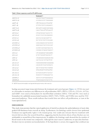
Table 1. Primer sequences used for RT-qPCR assays
Used pair 1
shEEF1A1-qF GGCACGTAGATTCAGGGAAG
shEEF1A1-qR CCCAGGCATATTTGAAGGAG
shKRT1-qF CTTCTGCAACCCCTCAATGT
shKRT1-qR GTTCTGCTGCTCCAGGAATC
shKRT10-qF GGTAATTCAAGCCAGCGAGA
shKRT10-qR CAGCCTGGCATTATCAACCT
shPCNA-qF CTTGGTGCAGCTAACCCTTC
shPCNA-qR ATGTCTTCATTGCCAGCACA
shTGFb1-qF GGGTACCACGCCAATTTCT
shTGFb1-qR GGTTGTGCTGGTTGTACAGG
shTGFBR1-qF CAACCAGGACCACTGCAATA
shTGFBR1-qR AAGCAGACTGGTCCAGCAAT
shTGFBR2-qF CCCTGTCGGTAGATGACCTG
shTGFBR2-qR CAGGGCCATGGAGTAGACAT
shVim-qF CTTCAGGAGGCTGAGGAATG
shVim-qR GTTGTTGCGGTTAGCAGCTT
shFN1-qF CTCGAAGAGCAGGAGACAGG
shFN1-qR CGCTCCCACTGTTGGTTTAT
shCol1a1-qF CAGGAAGAAGGCCAAGAAGA
shCol1a1-qR CACACGTCTCGGTCATGGTA
shActa2-qF AGCTATGAGCTGCCTGATGG
shActa2-qR GTACGTGGTCTCATGGATGC
shCol3a1-qF GGTGGACAGATTCTGGTGCT
shCol3a1-qR GGACATCTTCGGGAAGTTCA
shMMP1-qF AAATCCTCGTTGGGAGAACA
shMMP1-qR TTGGTCCACATCTGCTCTTG
RT-qPCR: Reverse transcription and quantitative polymerase chain reaction.
healing-associated target transcripts between the treatment and control groups [Figure 10]. PCNA was used
as a biomarker to measure any differences in cell proliferation. KRT1, KRT10, COL1A1, COL3A1, ACTA2,
and MMP1 were used as biomarkers for myofibroblast activation (MFA). VIM and FN1 were used as
biomarkers for epithelial-mesenchymal transition (EMT). TGF1, TGFR1, and TGFR2 were used for TGF-
associated pathways. These results indicate that timolol does not affect cell proliferation, at least at the
transcriptional level.
DISCUSSION
This study demonstrates that the topical application of timolol accelerates the epithelialization of mesh skin
grafted full-thickness burn wounds in sheep. Furthermore, the histology results showed that epidermal
thickness was significantly higher in the treated group. Our results also indicate that the treatment with
timolol did not affect the wound blood flow, suggesting that the beneficial effects of beta-blockers are not
attributable to wound blood flow improvement. In addition, the histology result showed that the number of
blood vessels in the wound was comparable. Although the exact reason is unknown, we speculate that beta-
blockers may not promote neovascularization. If the hypothesis is correct, this suggests that combined use