
Page 249 - Read Online
P. 249
Rice et al. Minocycline in spinal cord injury
results suggest that minocycline reduced microglia/
macrophage activation after injury.
We also determined the representation of microglia/
macrophages at the epicenter of injury. We found
that the density of Iba1 labeled cells was qualitatively
lower in minocycline treated mice (H) than in vehicle
controls (G), even though the morphology of cells,
with the majority being amoeboid, did not differ
between the 2 groups. To quantitate the extent of
microglial/macrophage reactivity encompassing
the lesion and remote areas, Iba1 immunoreactivity
was scored by three independent observers blinded
to treatment according to previously published
methods [12] . Agreement between observers was good
in large part and the identical result of 2 reviewers
was noted as the score for a particular section. The
blinded assessments [Figure 4I] indicated that there
was a significant difference in Iba1 immunoreactivity
between minocycline and vehicle treated mice 5 days
after injury (P < 0.05, Mann Whitney U test).
Minocycline decreases apoptotic cell death as
revealed by Tunel labeling
The number of Tunel positive cells was counted in
sections of spinal cords taken from mice at 2 and 5
days after injury. The data shows cell death occurring
at 2 and 5 days,and that minocycline treatment
reduced the number of Tunel positive cells at the latter
time point [Figure 5]. We did not address whether the
Tunel positive cells were oligodendrocytes and/or
neurons, and whether there is preferential rescue of
one cell type versus another by minocycline.
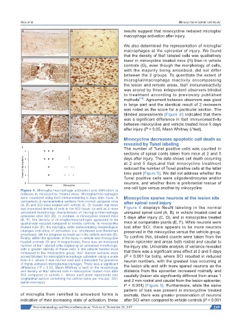
Figure 4: Microglial/macrophage activation and infiltration is
reduced in minocycline treated mice. Microglia/macrophages
were visualized using Iba1 immunostaining 5 days after injury. A Minocycline spares neurons at the lesion site
comparison of representative sections from normal uninjured mice after spinal cord injury
(A, B) and SCI mice treated with vehicle (C, D) reveals that there
was increased density of cells in the SCI tissue, as well as a more Figure 6 displays NeuN labeling in the normal
amoeboid morphology characteristic of microglial/macrophage uninjured spinal cord (A, B), in vehicle treated cord at
activation after SCI (D). In contrast, in minocycline treated mice 5 days after injury (C, D), and in minocycline treated
(E, F), the density of microglia/macrophages appeared to be
qualitatively reduced compared to vehicle controls. In minocycline mice at comparable points (E, F). While neurons were
treated mice (F), the microglia, while demonstrating morphological lost after SCI, there appears to be more neurons
changes indicative of activation (i.e. shortened and thickened preserved in the minocycline versus the vehicle group.
processes), did not progress as much as in the vehicle animals (D).
Finally, within the epicenter of the injury in vehicle and minocycline To confirm this, blinded counts were taken from the
treated animals (G and H respectively), there was an increased lesion epicenter and areas both rostral and caudal to
number of Iba1 labeled cells displaying an amoeboid morphology, the injury site. Univariate analysis of variance revealed
with a greater density of these cells in the vehicle treated mice
compared to the minocycline group. Iba1 stained sections were that there was a significant area effect at 2 and 5 days
scored blinded for microglial/macrophage activation using a scale (P < 0.001 for both), where SCI resulted in reduced
from 0-4, where 0 was normal cord and 4 indicated the presence neuron numbers, with the greatest loss occurring at
of highly activated microglia/macrophages. There was a significant the lesion site and with more spared neurons as the
difference (*P < 0.05, Mann Whitney U test) in the morphology
and density of Iba1 labeled cells in minocycline treated mice after distance from the epicenter increased rostrally and
SCI compared to vehicle (I, where each point represents one caudally (lesion site significantly different from areas 1
longitudinal section containing the central canal per mouse). SCI: and 2 mm rostral and caudal from the lesion epicenter,
spinal cord injury
P < 0.001) [Figure 6]. Furthermore, while the same
pattern of loss was present in minocycline treated
of microglia from ramified to amoeboid forms is animals, there was greater preservation of neurons
indicative of their increasing state of activation, these after SCI when compared to vehicle controls (P < 0.001
Neuroimmunology and Neuroinflammation ¦ Volume 4 ¦ November 28, 2017 249