
Page 226 - Read Online
P. 226
Figueroa-Hall et al. TLR4-mediated signaling in CHME-5 cells
A 4 B CHME-5 C 1.0
Ladder US LPS 0.8
3
kDa: 95
CD68 mRNA relative expression 2 72 CD68 CD68/β-tubulin 0.6
0.4
1
0.2
52 β-tubulin
0 0.0
US 3 6 18 h US LPS
LPS (μg/mL)
D CHME-5 NHA E 1.5
LPS (μg/mL) LPS (μg/mL)
CFAP/β-tubulin Fold Change 0.5
Ladder US 1 US 1 1.0
kDa: 72
GFAP
β-tubulin
52
0.0
US LPS US LPS
CHME-5 NHA
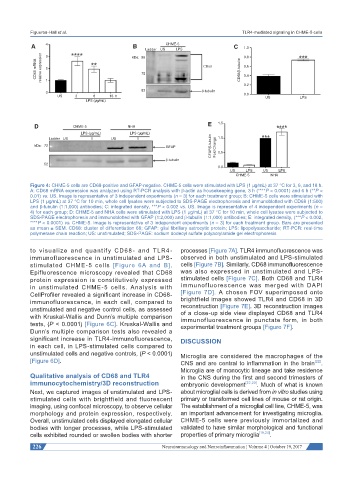
Figure 4: CHME-5 cells are CD68-positive and GFAP-negative. CHME-5 cells were stimulated with LPS (1 μg/mL) at 37 °C for 3, 6, and 18 h.
A: CD68 mRNA expression was analyzed using RT-PCR analysis with β-actin as housekeeping gene, 3 h (****P < 0.0001) and 6 h (**P <
0.01) vs. US. Image is representative of 3 independent experiments (n = 3) for each treatment group; B: CHME-5 cells were stimulated with
LPS (1 μg/mL) at 37 °C for 10 min, whole cell lysates were subjected to SDS-PAGE electrophoresis and immunoblotted with CD68 (1:500)
and β-tubulin (1:1,000) antibodies; C: integrated density, ***P < 0.002 vs. US. Image is representative of 4 independent experiments (n =
4) for each group; D: CHME-5 and NHA cells were stimulated with LPS (1 μg/mL) at 37 °C for 10 min, whole cell lysates were subjected to
SDS-PAGE electrophoresis and immunoblotted with GFAP (1:2,000) and β-tubulin (1:1,000) antibodies; E: integrated density, (***P < 0.002,
****P < 0.0001) vs. CHME-5. Image is representative of 3 independent experiments (n = 3) for each treatment group. Bars are presented
as mean ± SEM. CD68: cluster of differentiation 68; GFAP: glial fibrillary astrocytic protein; LPS: lipopolysaccharide; RT-PCR: real-time
polymerase chain reaction; US: unstimulated; SDS-PAGE: sodium dodecyl sulfate polyacrylamide gel electrophoresis
to visualize and quantify CD68 - and TLR4- processes [Figure 7A]. TLR4 immunofluorescence was
immunofluorescence in unstimulated and LPS- observed in both unstimulated and LPS-stimulated
stimulated CHME-5 cells [Figure 6A and B]. cells [Figure 7B]. Similarly, CD68 immunofluorescence
Epifluorescence microscopy revealed that CD68 was also expressed in unstimulated and LPS-
protein expression is constitutively expressed stimulated cells [Figure 7C]. Both CD68 and TLR4
in unstimulated CHME-5 cells. Analysis with immunofluorescence was merged with DAPI
CellProfiler revealed a significant increase in CD68- [Figure 7D]. A chosen FOV superimposed onto
immunofluorescence, in each cell, compared to brightfield images showed TLR4 and CD68 in 3D
unstimulated and negative control cells, as assessed reconstruction [Figure 7E]. 3D reconstruction images
of a close-up side view displayed CD68 and TLR4
with Kruskal-Wallis and Dunn’s multiple comparison immunofluorescence in punctate form, in both
tests, (P < 0.0001) [Figure 6C]. Kruskal-Wallis and experimental treatment groups [Figure 7F].
Dunn’s multiple comparison tests also revealed a
significant increase in TLR4-immunofluorescence, DISCUSSION
in each cell, in LPS-stimulated cells compared to
unstimulated cells and negative controls, (P < 0.0001) Microglia are considered the macrophages of the
[Figure 6D]. CNS and are central to inflammation in the brain [22] .
Microglia are of monocytic lineage and take residence
Qualitative analysis of CD68 and TLR4 in the CNS during the first and second trimesters of
immunocytochemistry/3D reconstruction embryonic development [22,23] . Much of what is known
Next, we captured images of unstimulated and LPS- about microglial cells is derived from in vitro studies using
stimulated cells with brightfield and fluorescent primary or transformed cell lines of mouse or rat origin.
imaging, using confocal microscopy, to observe cellular The establishment of a microglial cell line, CHME-5, was
morphology and protein expression, respectively. an important advancement for investigating microglia.
Overall, unstimulated cells displayed elongated cellular CHME-5 cells were previously immortalized and
bodies with longer processes, while LPS-stimulated validated to have similar morphological and functional
cells exhibited rounded or swollen bodies with shorter properties of primary microglia [19,24] .
226 Neuroimmunology and Neuroinflammation ¦ Volume 4 ¦ October 19, 2017