
Page 247 - Read Online
P. 247
Schindler et al. Microparticles in neuroimmune signaling
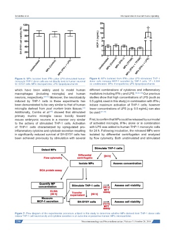
Figure 5: MPs isolated from IFN-γ plus LPS-stimulated human Figure 6: MPs isolated from IFN-γ plus LPS-stimulated THP-1
monocytic THP-1 donor cells are not directly toxic to human neuronal donor cells increase MCP-1 secretion by THP-1 cells. *P = 0.004
SH-SY5Y cells. MPs: microparticles; LPS: lipopolysaccharide vs. unstimulated. MPs: microparticles; LPS: lipopolysaccharide
which have been widely used to model human different combinations of cytokines and inflammatory
macrophages (including microglia) and human mediators including IFN-γ and LPS. [38,44,46] Our previous
neurons, respectively. [38-43] Moreover, the neurotoxicity studies show that high concentrations of LPS (such as
induced by THP-1 cells in these experiments has 0.5 μg/mL used in this study) in combination with IFN-γ
been demonstrated to be very similar to that of human induce maximum activation of THP-1 cells; however
microglia derived from post mortem brain tissues. lower concentrations of LPS (e.g. 0.5 ng/mL) can also
[44]
Additionally, Combs et al. showed that stimulated be used. [47,48]
[45]
primary murine microglia cause toxicity toward
mouse embryonic neurons in a manner very similar First, to confirm that MPs could be released by our model
to the actions of stimulated THP-1 cells. Activation of activated microglia, IFN-γ alone or in combination
of THP-1 cells characterized by upregulated pro- with LPS was added to human THP-1 monocytic cells
inflammatory cytokine and cytotoxin secretion resulting for 24 h. Following incubation, the released MPs were
in significantly reduced survival of SH-SY5Y cells has isolated by differential centrifugation and analyzed
been achieved previously by stimulation with several by flow cytometry. Both unstimulated and stimulated
Figure 7: Flow diagram of the experimental procedure utilized in this study to determine whether MPs derived from THP-1 donor cells
induce THP-1 cell neurotoxicity and cytokine secretion in an autocrine or paracrine manner. MPs: microparticles
238 Neuroimmunology and Neuroinflammation ¦ Volume 3 ¦ October 28, 2016