
Page 246 - Read Online
P. 246
Schindler et al. Microparticles in neuroimmune signaling
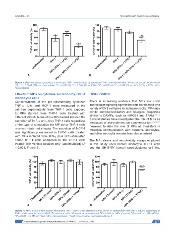
Figure 3: IFN-γ enhances cytotoxicity of monocytic THP-1 cells induced by stimulated THP-1 cell-derived MPs. *P = 0.034, 0.028 (A) *P = 0.021
#
#
&
&
(B) **P = 0.0001 (AB) vs. unstimulated; P = 0.002 (A) P = 0.004 (B) vs. IFN-γ; P = 0.0001(A) P = 0.007 (B) vs. MPs (IFN-γ + LPS). MPs:
microparticles
Effects of MPs on cytokine secretion by THP-1 DISCUSSION
monocytic cells
Concentrations of the pro-inflammatory cytokines There is increasing evidence that MPs are novel
TNF-α, IL-6, and MCP-1 were measured in the intercellular signaling agents that can be released by a
cell-free supernatants from THP-1 cells exposed variety of CNS cell types including microglia. MPs may
to MPs derived from THP-1 cells treated with exhibit immunomodulatory and biological properties
[13-16]
different stimuli. None of the MPs tested induced the similar to DAMPs, such as HMGB1 and TFAM.
secretion of TNF-α or IL-6 by THP-1 cells regardless Several studies have investigated the role of MPs as
mediators of astrocyte-neuron communication;
[15,16,27]
of the type of stimulation the MP donor THP-1 cells however, to date the role of MPs as mediators in
received (data not shown). The secretion of MCP-1 microglia communication with neurons, astrocytes,
was significantly enhanced in THP-1 cells treated and other microglia remains less characterized.
with MPs isolated from IFN-γ plus LPS-stimulated
donor THP-1 cells compared to the THP-1 cells The MP release and neurotoxicity assays employed
treated with vehicle solution only (unstimulated) (P in this study used human monocytic THP-1 cells
= 0.004, Figure 6). and the SH-SY5Y human neuroblastoma cell line,
Figure 4: MPs isolated from human monocytic THP-1 donor cells stimulated with TFAM in combination with IFN-γ induce cytotoxicity of
&
THP-1 cells towards human SH-SY5Y neuronal cells. *P = 0.01 vs. unstimulated; P = 0.004 vs. MPs (Control); P = 0.011 vs. MPs (IFN-γ);
#
% P = 0.0001 vs. MPs (TFAM). MPs: microparticles; TFAM: mitochondrial transcription factor A
Neuroimmunology and Neuroinflammation ¦ Volume 3 ¦ October 28, 2016 237