
Page 245 - Read Online
P. 245
Schindler et al. Microparticles in neuroimmune signaling
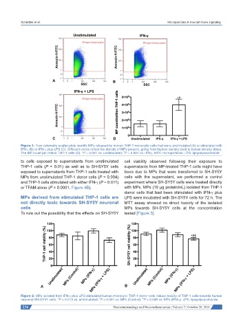
Figure 1: Flow cytometry scatter plots identify MPs released by human THP-1 monocytic cells that were unstimulated (A) or stimulated with
IFN-γ (B) or IFN-γ plus LPS (C). Different colors reflect the density of MPs present, going from highest density (red) to lowest density (blue).
#
The MP count per million THP-1 cells (D). *P = 0.001 vs. unstimulated; P = 0.001 vs. IFN-γ. MPs: microparticles; LPS: lipopolysaccharide
to cells exposed to supernatants from unstimulated cell viability observed following their exposure to
THP-1 cells (P = 0.01) as well as to SH-SY5Y cells supernatants from MP-treated THP-1 cells might have
exposed to supernatants from THP-1 cells treated with been due to MPs that were transferred to SH-SY5Y
MPs from unstimulated THP-1 donor cells (P = 0.004) cells with the supernatant, we performed a control
and THP-1 cells stimulated with either IFN-γ (P = 0.011) experiment where SH-SY5Y cells were treated directly
or TFAM alone (P = 0.0001, Figure 4B). with MPs. MPs (10 μg protein/mL) isolated from THP-1
donor cells that had been stimulated with IFN-γ plus
MPs derived from stimulated THP-1 cells are LPS were incubated with SH-SY5Y cells for 72 h. The
not directly toxic towards SH-SY5Y neuronal MTT assay showed no direct toxicity of the isolated
cells MPs towards SH-SY5Y cells at the concentration
To rule out the possibility that the effects on SH-SY5Y tested [Figure 5].
Figure 2: MPs isolated from IFN-γ plus LPS-stimulated human monocytic THP-1 donor cells induce toxicity of THP-1 cells towards human
#
neuronal SH-SY5Y cells. *P = 0.013 vs. unstimulated; P = 0.001 vs. MPs (Control); P = 0.048 vs. MPs (IFN-γ). LPS: lipopolysaccharide
&
236 Neuroimmunology and Neuroinflammation ¦ Volume 3 ¦ October 28, 2016