
Page 214 - Read Online
P. 214
Lira-Diaz et al. Emerging roles of microglia cells
epithelial layer located in the lateral walls of the lateral that migrate to the olfactory bulb through the rostral
ventricles. In the adult V-SVZ, three cell populations migratory stream and become mature interneurons.
[4]
have been identified: type-B cells, type-C cells and
type-A cells. The putative neural stem cell (NSC) is THE RELATIONSHIP BETWEEN
[5]
the type-B cell, an astroglial cell that can be identified MICROGLIA CELLS AND THE V-SVZ
by the expression of the glial fibrillary acidic protein,
glutamate aspartate transporter, brain lipid binding In the adult brain microglia cells are present all along
protein, platelet-derived growth factor receptor α, the V-SVZ [Figure 1] and remain in an intimate contact
CD133, Id1, Tailless, vascular cell adhesion molecule 1, with niche cells. The first interaction between the
[7]
epidermal growth factor receptor (EGFR), and others. [4,5] V-SVZ and microglia begins when microglia cells
The activation of B1 cells depends on signaling begin to populate the embryonic brain. At the early
pathways including sonic hedgehog, wingless-related stages of brain development an excessive number of
integration site, Notch, bone morphogenetic proteins, neurons are produced. This surplus of neurons needs
ephrins, retinoic acid, betacellulin, stromal derived to be eliminated and microglia cells are the responsible
factor-1, pigment epithelium-derived factor and some effectors of that function. Thus microglial cells are
intrinsic signals (Peroxiredoxin 1, sulfur oxide 2, crucial to maintain the balance of neurons and normal
arsenic-resistance 2, scute homolog 1, neuron-glia 2, postnatal brain development.
Oligodendrocyte lineage transcription factor 2). [4,5] After
activation, type-B1 cells produce transit-amplifying In the adult V-SVZ microglia cells stimulate
progenitors (type-C cells) that express the EGFR and neurogenesis by releasing soluble factors within
the transcription factors Dlx2 and Mash1. [5,6] Type-C the niche. This is a very complex process that
[5]
cells divide and give rise to neuroblasts (type-A cells) requires the molecular feedback between NSCs and
microglia cells, which release and express a myriad
of molecules, such as: CD200, vascular endothelial
growth factor, transforming growth factor β in NSCs
and CD200R, reactive nitrogen species/reactive
oxygen species, insulin-like growth factor 1, tumour
necrosis factor-α, Toll-like receptor-9, chemokine
fractalkine, chemokine fractalkine receptor (CX3CR1),
adenosine triphosphate-sensitive potassium channel
IL-1β, leukemia inhibitory factor, interferon-Ɣ. [7,8,9] In
the V-SVZ, microglia cells presents certain degree
of activation level and constantly release cytokines
and neurotrophic factors with respect to other brain
areas. This phenomenon suggests that NSCs are
regulated by microglia cells. All of these events occur
under physiological conditions, but under pathological
circumstances these signals can be magnified. After
activation, microglia cells enter into a phagocytic
state to remove cell debris and damaged. Phagocytic
microglia releases neurotrophic factors and cytokines
that activate NSCs, thus trigger cell survival and neural
regeneration after lesion. Microglial phagocytosis
is one of the principal mechanisms to regulate and
preserve the homeostasis in the production of neural
progenitors in the postnatal brain. Microglia also
eliminates aberrant cells that might later give rise to
malignant cells or brain tumors.
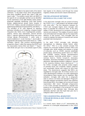
Figure 1: Microglia cells in the adult V-SVZ of mouse brain.
Schematic bran section: The adult V-SVZ is the neurogenic
niche lining the lateral walls of the lateral ventricles (LV). THE V-SVZ/ROSTRAL MIGRATORY
Photomicrography: Microglia cells labeled with anti-Iba1 antibodies STREAM MICROGLIA AND THEIR ROLE IN
and revealed with 3,3’-Diaminobenzidine (DAB) technique. At
resting stage, these cells exhibit ramified cell morphology and THE V-SVZ
numerous thin processes. Note that microglia cells are more
abundant in the V-SVZ as compared to adjacent brain regions:
[10]
corpus callosum (CC) and striatum (Str). These cells are also in In a recent report, Xavier et al. demonstrate the
close contact with blood vessels (BV). Bar = 20 µm presence of a microglial subpopulation in the V-SVZ
Neuroimmunology and Neuroinflammation ¦ Volume 3 ¦ September 26, 2016 205