
Page 160 - Read Online
P. 160
Interestingly, paw palpation similar to pain sensitivity body weight were measured every day. To assess
testing, and direct sensory nerve stimulation induce exploratory activity during the dark phase, animals
spinal transcription factor and IL‑1β expression, were placed in a dimly‑lit (10 Lux) open field device
respectively. [32‑34] In the present work, we therefore (40 cm × 40 cm) divided into 16 equal zones. Number
studied spinal and DRG expression as well as plasma of entries of the animal into a different zone and
concentrations of cytokines in murine models of arthritis rearing with or without leaning against the wall were
and bone cancer in relationship to signs of spontaneous scored during 10 min. [37] To study hind paw guarding
pain and paw palpation, rather than to pain sensitivity. during rearing, animals were introduced into an
inverted glass beaker of 20 cm diameter for 4 min
METHODS during the light phase. [38]
Animals As hallmarks of spinal neuroinflammation are variable
One hundred male C57/Bl6 mice(CharlesRiver,Arbresle, between studies, in particular among those using CFA,
France) weighing 25‑30 g and 46 male C3H/HeN we tested if mechanical non‑noxious stimulation is
mice (Janvier Labs, Le Genest St‑Isle, France) weighing one of the underlying factors. Therefore, half of the
20‑26 g were used. Four days before surgery, animals animals underwent hind paw palpation every second
were housed individually in plastic transparent cages for 2 min and were sacrificed 90 min later [Figure 1].
with unrestricted access to food and water in a room To avoid any effect of mechanical allodynia testing on
maintained at 21.5‑22.5 °C. Lights were on from 3:00 a.m. spinal gene expression, hind paw responses to von Frey
to 15:00 p.m. All experimental procedures were approved filaments (0.16‑2.4 g) applied to the plantar surface
by the local ethical committee (No. AP/2/11/2006). were studied a few minutes before sacrifice.
Arthritis and bone cancer induction Articular inflammation and bone destruction
C57/Bl6 mice were anesthetized with isoflurane and To assess inflammation, extracellular fluid was
placed in a supine position to insert a 26‑gauge needle detected in vivo using T2‑weighted magnetic resonance
[35]
into the knee joint as described by Gauldie et al. Fifty µL imaging (MRI) on a 4.7 T horizontal magnet (Bruker,
of complete Freund adjuvant (CFA; Sigma‑Aldrich, Ettlingen, Germany). To determine bone destruction,
St. Louis, MO, USA) or mineral oil vehicle were 3D FLASH‑based magnetic resonance microscopy
injected on days 0 and 6 into the same joint [Figure 1]. of femurs was carried out ex vivo on a 9.4 T vertical
C3H/HeN mice were anesthetized with isoflurane and magnet (Bruker Biospec 47/50, Ettlingen, Germany).
injected with 10 µL of phosphate buffer saline (PBS)
5
containing 10 NCTC‑2472 cells (LGC Promochem, Tissue preparation
Molsheim, France) propagated in vitro or 10 µL of Two days after the second intra‑articular injection, that
PBS into the intramedullary canal of the femur in is 8 days after the first injection, or 21 days after femur
accordance with a previous report by Schwei et al. [33] injection [Figure 1], animals were deeply anesthetized
with sodium pentobarbital to allow for intracardiac
Behavioral testing puncture. Animals were rinsed with PBS after which
Reduced food intake and exploration are signs of animals assigned to immunohistochemical analysis
pain in rodents. [36] After surgery, food pellets and were perfused with 4% paraformaldehyde in 0.1 mol/L
PBS. L3‑L5 spinal cords and DRGs of these animals were
Hind Paw post‑fixed for 4 h, cryoprotected in 30% sucrose, frozen
palpation
Open Field Beaker Open Field Beaker Open Field OR on dry ice and stocked at ‑80 °C. For animals allocated
Session 1 Session 1 Session 2 Session 2 Session 3 Von Frey to polymerase chain reaction (PCR) experiments, L3‑L5
Time spinal cords and DRGs were removed within 3 min after
D0 D1 D2 D4 D5 D6 D7 D8 rinsing with PBS and then frozen at ‑80 °C.
First CFA Second CFA MRI Sacrifice
injection injection Spinal Fos expression
a Beaker Immunohistochemical detection of c‑Fos and FosB
Session 2
Open Field Open Field Beaker Open Field transcription factors in the spinal cord was performed
Session 1 Session 2 Session 1 Session 3 Von Frey using rabbit antisera (diluted 1:2000; Santa Cruz
Time Biotechnology, Santa Cruz, CA, USA) as previously
D0 D14 D17 D18 D20 D21 D22 described. [39]
MRI
Tumor cell Sacrifice
b injection Circulating cytokines
Blood samples were collected in EDTA‑coated vials,
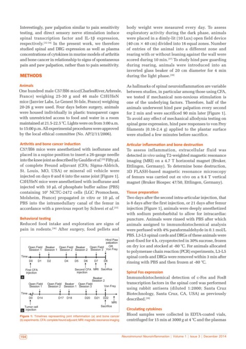
Figure 1: Timelines representing joint inflammation (a) and bone cancer
(b) experiments. CFA: complete freund adjuvant; MRI: magnetic resonance imaging centrifuged for 15 min at 3000 g at 4 °C and the plasmas
154 Neuroimmunol Neuroinflammation | Volume 1 | Issue 3 | December 2014