
Page 155 - Read Online
P. 155
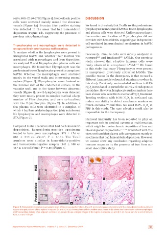
(42%; 95% CI: 20‑67%) [Figure 1]. Hemosiderin positive   DISCUSSION
           cells were scattered mainly around the abnormal
           vessels [Figure 1a]. Prussian blue positive staining   We found in this study that T‑cells are the predominant
           was  detected  in  the  areas  that  had  hemosiderin   lymphocytes in unruptured bAVMs. Few B‑lymphocytes
           deposition  [Figure  1d], suggesting the presence of   and plasma cells were detected. Unlike macrophages,
           previous micro‑hemorrhage.                         the number and location of T‑lymphocytes did not
                                                              correlate with hemosiderin, suggesting an independent
                                                              cell‑mediated immunological mechanism in bAVM
           T-lymphocytes and macrophages were detected in     pathogenesis.
           unruptured brain arteriovenous malformations
           To analyze whether the lymphocytes were present in   Previously, immune cells were mostly analyzed in
           unruptured bAVM and whether their location was     ruptured [24]  and irradiated [25]  bAVMs. Our previous
           associated with macrophages and iron deposition,   study  showed  that  adaptive  immune  cells  were
           we analyzed T‑ and B‑lymphocytes, plasma cells and   rarely  observed  in  unruptured  bAVM. [13]   We  found
           macrophages. We found that T‑lymphocyte was the    in this study that many T‑lymphocytes were present
           predominant type of lymphocytes present in unruptured   in unruptured, previously untreated bAVMs. The
           bAVM. Whereas the macrophages were scattered       possible reason for the discrepancy is that we used a
           mostly in the vessel walls and intervening stromal   different immunohistochemical staining procedure in
           regions [Figure 2], T‑lymphocytes were clustered on   this study. Previously, we incubated sections in 0.3%
           the  luminal  side  of  the  endothelial  surface,  in  the   H O  in methanol to quench the activity of endogenous
           vascular wall, and in the tissue between abnormal   peroxidase. However, lymphocyte surface markers have
                                                                2
                                                                  2
           vessels [Figure 3]. Few B‑lymphocytes were detected;   been shown to be sensitive to methanol/H O  treatment.
           they were mostly present in samples that had a large   Treating sections with 0.3% H O  in methanol can
                                                                                                    2
                                                                                                  2
           number  of  T‑lymphocytes,  and  were  co‑localized   reduce our ability to detect membrane markers on
                                                                                             2
                                                                                           2
           with the T‑lymphocytes  [Figure  2]. In addition, a   frozen sections, [26]  and thus, we used 0.3% H O  in
           few plasma cells were identified in 5  samples, of   PBS in this study. The case selection could also be
                                                                                                           2
                                                                                                         2
           which 4 had hemosiderin deposition (data not shown).   responsible for the discrepancy.
           No lymphocytes and macrophages were detected in
           STA [Figure 2].
                                                              Humoral immunity has been reported to play an
                                                              important role in cerebral cavernous malformation,
           Compared to the specimens that had no hemosiderin   which might be due to chronic deposition of iron and
           deposition,  hemosiderin‑positive  specimens       blood degradation products. [22,27,28]  Consistent with this
           tended to have more macrophages  (478  ±  174  vs.   view, we found that plasma cells were present mainly in
           666  ±  313  cells/mm ; P  =  0.11). The T‑cell    specimens that had hemosiderin deposition. However,
                                 2
           numbers were similar in hemosiderin‑positive       we cannot draw any conclusion regarding adaptive
           and hemosiderin‑negative samples  (147  ±  108  vs.   immune responses to the presence of iron from our
           157 ± 139 cells/mm ; P = 0.88) [Figure 4].         small descriptive study.
                             2


                             a                      b                      c


                             d                      e                      f
           Figure 1: Hemosiderin deposition in unruptured brain arteriovenous malformations. H and E (a‑c) and prussian blue staining (d‑f) on the adjacent sections. (b) and
           (c) are enlarged pictures of the regions in squares (b) and (c) in (a) showing hemosiderin‑positive areas. Insert in (b) shows two hemosiderin‑laden macrophages.
           (d) Prussian blue staining of an adjacent section of (a). (e) and (f) are enlarged images of the regions in squares (e) and (f) in (d). Scale bars for a and d: 500 μm;
           for b, c, e and f: 50 μm


          Neuroimmunol Neuroinflammation | Volume 1 | Issue 3 | December 2014                               149