
Page 96 - Read Online
P. 96
Ackerman et al. Mini-invasive Surg 2021;5:14 https://dx.doi.org/10.20517/2574-1225.2021.02 Page 15 of 19
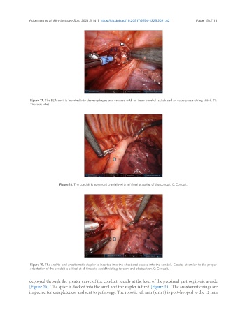
Figure 17. The EEA anvil is inserted into the esophagus and secured with an inner baseball stitch and an outer purse-string stitch. TI:
Thoracic inlet.
Figure 18. The conduit is advanced cranially with minimal grasping of the conduit. C: Conduit.
Figure 19. The end-to-end anastomotic stapler is inserted into the chest and passed into the conduit. Careful attention to the proper
orientation of the conduit is critical at all times to avoid twisting, torsion, and obstruction. C: Conduit.
deployed through the greater curve of the conduit, ideally at the level of the proximal gastroepiploic arcade
[Figure 20]. The spike is docked into the anvil and the stapler is fired [Figure 21]. The anastomotic rings are
inspected for completeness and sent to pathology. The robotic left arm (arm 1) is port-hopped to the 12 mm