
Page 97 - Read Online
P. 97
Page 16 of 19 Ackerman et al. Mini-invasive Surg 2021;5:14 https://dx.doi.org/10.20517/2574-1225.2021.02
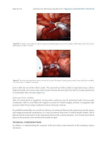
Figure 20. The end-to-end anastomotic spike is deployed through the greater curve of the conduit, ideally at the level of the proximal
gastroepiploic arcade. C: Conduit.
Figure 21. The end-to-end anastomotic spike is docked into its anvil. The stapler is gently approximated, closed, and fired to complete
the anastomosis. C: Conduit; E: esophagus.
port to allow for use of the robotic stapler. The proximal tip of the conduit is amputated using a robotic
stapler load with care to leave some tissue distance between the new staple line and the circular anastomosis
to avoid undue tissue ischemia [Figure 22].
Endoscopy, drains, and flaps
After the anastomosis is completed, intraoperative endoscopy may be performed under thoracoscopic
visualization with the chest filled with irrigation to assess for conduit integrity and leak. A nasogastric tube
is passed under thoracoscopic visualization before endoscope removal.
If a pedicled omental flap was created (not shown), it is interposed between the anastomosis and the airway,
and wrapped around the anastomosis. A 10 mm flat Jackson-Pratt drain (Cardinal Health, Dublin, OH) is
placed adjacent and posterior to the anastomosis between the conduit and spine. A 28-French chest tube is
placed in the posterior chest and directed towards the apex.
TECHNICAL CONSIDERATIONS
In addition to understanding the sequence of the procedure, some elements of the technique require
discussion.