
Page 95 - Read Online
P. 95
Page 14 of 19 Ackerman et al. Mini-invasive Surg 2021;5:14 https://dx.doi.org/10.20517/2574-1225.2021.02
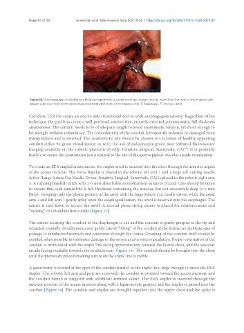
Figure 16. The esophagus is divided at a level appropriate for a sound oncologic margin, but no lower than the level of the azygous vein.
Shown is division high in the chest at approximately the level of the thoracic inlet. E: Esophagus; TI: thoracic inlet.
Covidien, USA) to create an end-to-side (functional end-to-end) esophagogastrostomy. Regardless of the
technique, the goal is to create a well-perfused, tension-free, properly oriented, pneumostatic, full-thickness
anastomosis. The conduit needs to be of adequate length to avoid anastomotic tension, yet short enough to
lay straight without redundancy. The redundant tip of the conduit is frequently ischemic or damaged from
manipulation and is resected. The anastomotic site should be chosen at a location of healthy appearing
conduit either by gross visualization or with the aid of indocyanine green near-infrared fluorescence
[30]
imaging available on the robotic platform (Firefly, Intuitive Surgical, Sunnyvale, CA) . It is generally
feasible to create the anastomosis just proximal to the site of the gastroepiploic vascular arcade termination.
To create an EEA stapled anastomosis, the stapler anvil is inserted into the chest through the anterior aspect
of the access incision. The Force Bipolar is placed in the robotic left arm 1 and a large self-cutting needle
driver (Large Suture Cut Needle Driver, Intuitive Surgical, Sunnyvale, CA) is placed in the robotic right arm
3. A running baseball stitch with 2-0 non-absorbable monofilament suture is placed. Care should be taken
to ensure that each suture bite is full-thickness containing the mucosa, but not excessively deep (2-3 mm
bites). Grasping only the plastic portion of the anvil with the large Suture Cut needle driver, while the assist
arm 4 and left arm 1 gently splay open the esophageal lumen, the anvil is inserted into the esophagus. The
suture is tied down to secure the anvil. A second purse-string suture is placed for reinforcement and
“tucking” of redundant tissue folds [Figure 17].
The suture securing the conduit to the diaphragm is cut and the conduit is gently grasped at the tip and
retracted cranially. Simultaneous and gentle lateral “lifting” of the conduit at the hiatus can facilitate ease of
passage of tubularized stomach and omentum through the hiatus. Grasping of the conduit itself should be
avoided when possible to minimize damage to the serosa and/or microvasculature. Proper orientation of the
conduit is maintained with the staple line facing approximately towards the lateral chest, and the vascular
arcade facing medially towards the mediastinum [Figure 18]. The conduit should be brought into the chest
until the previously placed marking suture on the staple line is visible.
A gastrotomy is created at the apex of the conduit parallel to the staple line, large enough to insert the EEA
stapler. The robotic left arm and port are removed, the conduit is oriented toward the access incision, and
the conduit lumen is irrigated with antibiotic-infused saline. The EEA stapler is inserted through the
anterior portion of the access incision along with a laparoscopic grasper and the stapler is passed into the
conduit [Figure 19]. The conduit and stapler are brought together into the upper chest and the spike is