
Page 91 - Read Online
P. 91
Page 10 of 19 Ackerman et al. Mini-invasive Surg 2021;5:14 https://dx.doi.org/10.20517/2574-1225.2021.02
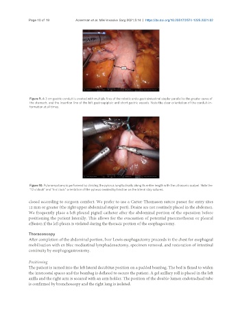
Figure 9. A 3 cm gastric conduit is created with multiple fires of the robotic endo-gastrointestinal stapler parallel to the greater curve of
the stomach, and the insertion line of the left gastroepiploic and short gastric vessels. Note the clear orientation of the conduit-in-
formation at all times.
Figure 10. Pyloromyotomy is performed by dividing the pylorus longitudinally along its entire length with the ultrasonic scalpel. Note the
“12 o’clock” and “6 o’clock” orientation of the pylorus created by traction on the lateral stay sutures.
closed according to surgeon comfort. We prefer to use a Carter-Thomason suture passer for entry sites
12 mm or greater (the right upper abdominal stapler port). Drains are not routinely placed in the abdomen.
We frequently place a left pleural pigtail catheter after the abdominal portion of the operation before
positioning the patient laterally. This allows for the evacuation of potential pneumothorax or pleural
effusion if the left pleura is violated during the thoracic portion of the esophagectomy.
Thoracoscopy
After completion of the abdominal portion, Ivor Lewis esophagectomy proceeds in the chest for esophageal
mobilization with en bloc mediastinal lymphadenectomy, specimen removal, and restoration of intestinal
continuity by esophagogastrostomy.
Positioning
The patient is turned into the left lateral decubitus position on a padded beanbag. The bed is flexed to widen
the intercostal spaces and the beanbag is deflated to secure the patient. A gel axillary roll is placed in the left
axilla and the right arm is secured with an arm holder. The position of the double-lumen endotracheal tube
is confirmed by bronchoscopy and the right lung is isolated.