
Page 89 - Read Online
P. 89
Page 8 of 19 Ackerman et al. Mini-invasive Surg 2021;5:14 https://dx.doi.org/10.20517/2574-1225.2021.02
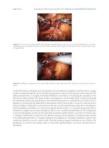
Figure 4. The celiac axis is skeletonized along the left gastric vascular pedicle, splenic artery, and common hepatic artery. All lymph
node bearing tissue is dissected, elevated, and kept with the specimen. LGA: Left gastric artery and vein; CHA: common hepatic artery;
SA: splenic artery.
Figure 5. The left gastric artery and vein are divided with a robotic vascular staple load. LGAV: Left gastric artery and vein; SA: splenic
artery.
reaches the anterior abdominal wall. An insertion site in the left lower quadrant is selected and a 25-gauge
needle is inserted through the skin to identify the jejunostomy tube site. The jejunum is then sutured to the
abdominal wall with a 2-0 Surgidac Endostitch (Medtronic, New Haven, CT), keeping the orientation of the
afferent and efferent limbs. The finder needle is exchanged for a Yueh needle, which is advanced through
the abdominal wall into the jejunal lumen. The intraluminal position is confirmed by an air bolus. Next, a
guidewire is inserted into the distal limb of the jejunum and the Yueh needle is removed. A skin incision is
made, the dilator and sheath are advanced over the wire into the jejunal lumen under direct visualization,
and the guidewire and dilator are removed leaving the sheath in place. A 10-French jejunostomy tube is
trimmed to a length of 20 cm from the balloon, which is cut to avoid accidental inflation. The feeding tube
is advanced through the sheath into the distal limb of jejunum and the sheath is removed. Two Witzel-type
2-0 Surgidac Endostitches are placed on the efferent jejunum and the jejunum is circumferentially sutured
to the abdominal wall with a 2-0 Surgidac Endostitch. An additional 2-0 Surgidac Endostitch is placed a few
centimeters distally as an anti-torsion stitch. The tube position is again confirmed by an air bolus. The
feeding tube is secured to the bumper with a 2-0 silk suture and the bumper is secured to the skin with 2-0
silk sutures.