
Page 88 - Read Online
P. 88
Ackerman et al. Mini-invasive Surg 2021;5:14 https://dx.doi.org/10.20517/2574-1225.2021.02 Page 7 of 19
Figure 2. Early circumferential hiatal mobilization is performed to assess the extent of local disease and ensure resectability. E:
Esophagus; RC: right crural pillar; LC: left crural pillar; Ao: aorta.
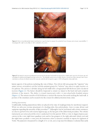
Figure 3. Retrogastric exposure is obtained by retracting the stomach anteriorly by passing the robotic assist arm into the lesser sac and
elevating the gastroesophageal junction anteriorly. The bedside assist arm provides further retraction of the stomach and lesser
omentum, as shown, to clearly expose the vascular pedicle and retrogastric space. LGAV: Left gastric artery and vein.
lateral aspects of the pylorus including the vein of Mayo. The robotic left arm grasps the “superior” stay
suture (screen orientation) and the bedside assistant grasps the “inferior” stay suture to apply traction on
the pylorus. The pylorus is divided along its full width with a longitudinal full-thickness antro-duodenal
incision [Figure 10]. The lumen should be inspected to ensure no injury to the back wall and complete
division of the muscle. The defect is closed transversely with 2-0 non-absorbable braided sutures
[Figure 11]. The sutures must be of full-thickness to include the mucosa, but avoid catching the back wall. A
portion of the omentum can be secured over the closure for added protection from a leak.
Feeding jejunostomy
Traditionally, feeding jejunostomy tubes are placed at the time of esophagectomy for nutritional support.
While we advocate routine placement of a feeding tube, like pyloroplasty, there is some debate and
equipoise regarding the necessity of this procedure . Although it can be performed with robotic assistance,
[28]
we elect to perform this with routine laparoscopy as the last part of the abdominal procedure. The robotic
instruments are removed and the robot is undocked from the patient. With a 5 mm 30-degree laparoscopic
camera in the 5 mm right lower quadrant port, and in-line graspers in the right subcostal robotic port and
the right lower quadrant 12 mm port, the transverse colon is retracted cranially to expose the ligament of
Treitz. The jejunum is measured for a length of 35-40 cm, and a loop near this distance is selected that easily