
Page 86 - Read Online
P. 86
Ackerman et al. Mini-invasive Surg 2021;5:14 https://dx.doi.org/10.20517/2574-1225.2021.02 Page 5 of 19
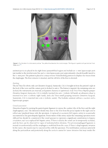
Figure 1. Port location for abdominal portion. The yellow line denotes the costal margin. This figure is quoted with permission from
[27]
Ekeke et al. .
assistant ports are placed in the right lower paraumbilical region and include an 11 mm laparoscopic port
just medial to the midclavicular line and a 5 mm laparoscopic port approximately a hands breadth lateral to
the 11 mm port. The patient is placed in a steep reverse-Trendelenburg position to displace the viscera from
the diaphragm. The liver retractor is inserted, and the left lobe of the liver is elevated.
Docking
The da Vinci Xi robotic side cart (Intuitive Surgical, Sunnyvale, CA) is brought in from the patient’s right at
the level of the torso and the camera port is docked to arm 2. The hiatus is targeted, the remaining arms are
docked, the instruments are inserted, and patient clearance is optimized. A da Vinci Force Bipolar grasper
(Intuitive Surgical, Sunnyvale, CA) is initially inserted into arm 1 (robotic left hand), an ultrasonic shear is
inserted into arm 3 (robotic right hand), and a da Vinci small grasping retractor (Intuitive Surgical,
Sunnyvale, CA) is inserted into arm 4 (robotic assist). The bedside assistant utilizes a suction and a
laparoscopic grasper.
Crural assessment
Dissection begins by excising the gastrohepatic ligament to expose the caudate lobe of the liver and the right
diaphragmatic crus. The dissection should stay close to the liver from the porta hepatis to the right crus to
reflect any lymphoid tissue with the specimen. A replaced or accessory left hepatic artery is occasionally
encountered in the gastrohepatic ligament. Preservation of this artery makes the remaining operation more
difficult but should be considered if the vessel appears to represent a significant contribution to hepatic
circulation, such as a replaced left hepatic artery. If there is doubt, the vessel can be temporarily occluded,
and the liver can be observed for signs of ischemia before division. The phrenoesophageal ligament is
incised circumferentially and the esophagus is mobilized from the crura. If there is diaphragmatic invasion
by the tumor, the muscle may be resected en bloc with the specimen. The mobilization continues anteriorly
along the pericardium and posteriorly along the aorta to assess for tumor invasion that may render the