
Page 166 - Read Online
P. 166
Tian et al. EMT drives anti-estrogen resistance in breast cancer
also significantly more sensitive to growth inhibition [Figure 5C and D] assays. Taken together, these results
by administration of small molecule antagonists to demonstrated that post-EMT cells acquire resistance
either IGF1R (i.e. AG1024; Supplementary Figure 5A) to tamoxifen by upregulating EGFR and IGF1R
or EGFR (i.e. AG1478; Supplementary Figure 5B), expression and MAP kinase activation, culminating in
findings consistent with the ability of post-EMT cells to extranuclear localization and nongenomic signaling of
upregulate their expression of IGF1R and EGFR and ER-α in MCF-7 cells.
activation of ERK1/2 [Figures 3 and 4]. Additionally,
post-EMT MCF-7 cells also exhibited significantly DISCUSSION
increased cell growth and decreased sensitivity to
tamoxifen-induced cell death [Figure 5B]. Importantly, The induction of EMT programs by TGF-β plays
co-administration of tamoxifen with small molecule important roles in driving the progression, dissemination,
inhibitors against either TβR-I (i.e. TβR-I inhibitor II), and recurrence of human breast cancers; these events
IGF1R (i.e. AG1024), EGFR (i.e. AG1478), or MEK1/2 also underlie the development, expansion, and self-
(i.e. U0126) restored MCF-7 cell sensitivity to tamoxifen renewal of cancer stem cells, as well as the acquisition
as determined by MTS [Figure 5B] or clonogenic of chemoresistant phenotypes. [4,22,23] Although EMT
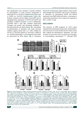
Figure 5: TGF-β stimulation of EMT programs promotes tamoxifen resistance in MCF-7 cells. (A) Pre- and post-EMT MCF-7 organoids
were treated with estradiol (0.1 nmol/L), tamoxifen (0.1 µmol/L), or fulvestrant (0.1 µmol/L) for 8 days, at which point photomicrographs
were captured and analyzed on Image J to assess differences in organoid growth. Images are representative of 3-independent
experiments, while data are the mean fold-changes (± SE; n = 3; *P < 0.05; Student’s t-test; ×400); (B) pre- and post-EMT MCF-7 cells
were treated with tamoxifen (0.1 µmol/L) in the absence or presence of TβR-I Inhibitor II (100 ng/mL; left), of AG1024 (1 µmol/L; middle),
or of AG1478 (1 µmol/L; right) as indicated. Afterward, differences in cell growth and survival were analyzed by MTS assays. Data are
the mean (± SE; n = 3; *P < 0.05; Student’s t-test) growth relative to untreated pre-EMT cells (*P < 0.05; Student’s t-test); (C-E) pre- and
post-EMT MCF-7 cells were treated with tamoxifen (0.1 µmol/L) in the absence or presence of U0126 (10 µmol/L; left), of TβR-I Inhibitor
II (100 ng/mL; middle), or AG1024 (1 µmol/L; right) for 10 days, at which point the number of surviving colonies in 11 random fields/
plate was enumerated. Data are the mean (± SE; n = 3; *P < 0.05; Student’s t-test). TGF: transforming growth factor; EMT: epithelial-
mesenchymal transition; SE: standard error
158 Journal of Cancer Metastasis and Treatment ¦ Volume 3 ¦ August 21, 2017