
Page 164 - Read Online
P. 164
Tian et al. EMT drives anti-estrogen resistance in breast cancer
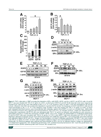
Figure 3: TGF-β stimulation of EMT promotes the interaction of ER-α with EGFR, IGF1R, and Src in MCF-7 and BT474 cells. (A and B)
MCF-7 cells were treated with TGF-β1 (5 ng/mL) for 0-96 h, at which point EGFR (A) and IGF1R (B) transcript levels were measured by
real-time PCR. Data are the mean fold-changes (± SE; n = 3; *P < 0.05; Student’s t-test). (C) MCF-7 cells were propagated in 2D and 3D
cultures. Afterward, total RNA was isolated and subjected to real-time PCR to monitor the expression of EGFR and IGF1R. Data are the
mean fold-changes (± SE; n = 3; *P < 0.05; Student’s t-test); (D) MCF-7 cells were treated with TGF-β1 (5 ng/mL) for 0-96 h, at which
point detergent-solubilized extracts were immunoblotted with antibodies against either EGFR or β-actin as indicated (top). Additionally,
ER-α immunocomplexes were captured and immunoblotted for EGFR and ER-α as indicated (bottom). Images are representative of
4-independent experiments; (E) BT474 cells were stimulated with TGF-β1 (5 ng/mL) as indicated, and subsequently were subjected to
immunoblot analyses to monitor the expression of EGFR, IGF1R, and β-actin. Images are representative of 3-individual experiments;
(F and G) BT474 cells were stimulated with TGF-β1 (5 ng/mL) for 0-72 h, while MCF-7 cells were stimulated with TGF-β1 for 0-96 h.
Afterward, detergent-solubilized extracts were immunoprecipitated with antibodies against ER-α and subsequently were immunoblotted
with antibodies against IGF1R and ER-α. Images are representative of at least 3-individual experiments; (H) MCF-7 cells were treated
with TGF-β1 (5 ng/mL) for 0-96 h, at which point detergent-solubilized extracts were immunoblotted for Src or β-actin as indicated (top).
Additionally, Src immunocomplexes were captured and immunoblotted for ER-α and Src as indicated (bottom). Images are representative
of 4-independent experiments. TGF: transforming growth factor; EMT: epithelial-mesenchymal transition; IGF: insulin-like growth factor; ER:
estrogen receptor; EGFR: epidermal growth factor receptor; SE: standard error
156 Journal of Cancer Metastasis and Treatment ¦ Volume 3 ¦ August 21, 2017