
Page 165 - Read Online
P. 165
Tian et al. EMT drives anti-estrogen resistance in breast cancer
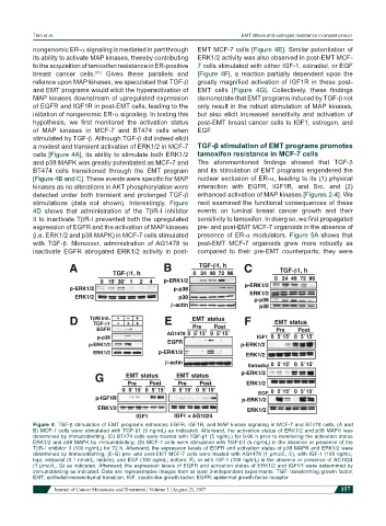
nongenomic ER-α signaling is mediated in part through EMT MCF-7 cells [Figure 4E]. Similar potentiation of
its ability to activate MAP kinases, thereby contributing ERK1/2 activity was also observed in post-EMT MCF-
to the acquisition of tamoxifen resistance in ER-positive 7 cells stimulated with either IGF-1, estradiol, or EGF
breast cancer cells. Given these parallels and [Figure 4F], a reaction partially dependent upon the
[31]
reliance upon MAP kinases, we speculated that TGF-β greatly magnified activation of IGF1R in these post-
and EMT programs would elicit the hyperactivation of EMT cells [Figure 4G]. Collectively, these findings
MAP kinases downstream of upregulated expression demonstrate that EMT programs induced by TGF-β not
of EGFR and IGF1R in post-EMT cells, leading to the only result in the robust stimulation of MAP kinases,
initiation of nongenomic ER-α signaling. In testing this but also elicit increased sensitivity and activation of
hypothesis, we first monitored the activation status post-EMT breast cancer cells to IGF1, estrogen, and
of MAP kinases in MCF-7 and BT474 cells when EGF.
stimulated by TGF-β. Although TGF-β did indeed elicit
a modest and transient activation of ERK1/2 in MCF-7 TGF-β stimulation of EMT programs promotes
cells [Figure 4A], its ability to stimulate both ERK1/2 tamoxifen resistance in MCF-7 cells
and p38 MAPK was greatly potentiated as MCF-7 and The aforementioned findings showed that TGF-β
BT474 cells transitioned through the EMT program and its stimulation of EMT programs engendered the
[Figure 4B and C]. These events were specific for MAP nuclear exclusion of ER-α, leading to its (1) physical
kinases as no alterations in AKT phosphorylation were interaction with EGFR, IGF1R, and Src, and (2)
detected under both transient and prolonged TGF-β enhanced activation of MAP kinases [Figures 2-4]. We
stimulations (data not shown). Interestingly, Figure next examined the functional consequences of these
4D shows that administration of the TβR-I inhibitor events on luminal breast cancer growth and their
II to inactivate TβR-I prevented both the upregulated sensitivity to tamoxifen. In doing so, we first propagated
expression of EGFR and the activation of MAP kinases pre- and post-EMT MCF-7 organoids in the absence of
(i.e. ERK1/2 and p38 MAPK) in MCF-7 cells stimulated presence of ER-α modulators. Figure 5A shows that
with TGF-β. Moreover, administration of AG1478 to post-EMT MCF-7 organoids grew more robustly as
inactivate EGFR abrogated ERK1/2 activity in post- compared to their pre-EMT counterparts; they were
Figure 4: TGF-β stimulation of EMT programs enhances EGFR, IGF1R, and MAP kinase signaling in MCF-7 and BT474 cells. (A and
B) MCF-7 cells were stimulated with TGF-β1 (5 ng/mL) as indicated. Afterward, the activation status of ERK1/2 and p38 MAPK was
determined by immunoblotting; (C) BT474 cells were treated with TGF-β1 (5 ng/mL) for 0-96 h prior to monitoring the activation status
ERK1/2 and p38 MAPK by immunoblotting. (D) MCF-7 cells were stimulated with TGF-β1 (5 ng/mL) in the absence or presence of the
TβR-I inhibitor II (100 ng/mL) for 72 h. Afterward, the expression levels of EGFR and activation status of p38 MAPK and ERK1/2 were
determined by immunoblotting; (E-G) pre- and post-EMT MCF-7 cells were treated with AG1478 (1 µmol/L; E), with IGF-1 (100 ng/mL;
top), estradiol (0.1 nmol/L; middle), and EGF (100 ng/mL; bottom; F), or with IGF-1 (100 ng/mL) in the absence or presence of AG1024
(1 µmol/L; G) as indicated. Afterward, the expression levels of EGFR and activation status of ERK1/2 and IGF1R were determined by
immunoblotting as indicated. Data are representative images from at least 3-independent experiments. TGF: transforming growth factor;
EMT: epithelial-mesenchymal transition; IGF: insulin-like growth factor; EGFR: epidermal growth factor receptor
Journal of Cancer Metastasis and Treatment ¦ Volume 3 ¦ August 21, 2017 157