
Page 47 - Read Online
P. 47
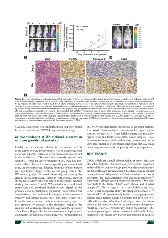
Figure 6: In vivo validation of withaferin A-mediated anti-cancer effects. (a) Representative bioluminescence images of control and withaferin A-treatment
            mice subcutaneously inoculated with floating (F) cells. Withaferin A treatment was initiated 2 weeks post-tumor implantation to allow tumor establishment.
            Note, inoculated F cells were found to start disseminating anteriorly during week 3-4 as compared to localized signal found in withaferin A-treated mice (left
            panels). The bioluminescent intensity was significantly lower in withaferin A-treated group than in the control group. The data were quantitatively represented
            by the fold change in bioluminescence intensity over time (right panel); (b) systematic injection of F cells mimicking metastatic model. Representative images
            of F cell-inoculated mice (via tail vein injection) demonstrated that withaferin A treatment given 2 weeks after intervenous tumor injection not only suppressed
            tumor growth but also controlled the spread of F cells; (c) immunohistochemical analysis of tumor samples harvested from both control and withaferin A-treated
            animals (from subcutaneous tumor samples). Representative sections of the tumor samples were stained with CXCR4, caspase-3, and poly ADP-ribose
            polymerase antibodies. Samples treated with withaferin A demonstrated decreased CXCR4 immunostaining, while increased caspase-3 and poly ADP-ribose
            polymerase immunostaining as compared with the control samples (×200)
            CXCL12 expression. The similarity in the response profile   of CXCR4 was significantly less intense in the tumor sections
            between vimentin and CXCR4 expression is striking.  from WA-treated mice than in control samples [Figure 6c]. In
                                                               contrast, caspase-3, -8, -9 and PARP staining was markedly
            In vivo validation of WA-mediated suppression      higher in the WA-treated xenografted tumor samples. These
            of tumor growth and metastasis                     in vivo observations, while preliminary, corroborated our in
                                                               vitro data (induction of apoptosis), suggesting that WA could
            Finally,  we  wished  to  validate  the  anti-cancer  effects   indeed suppress metastatic propensity and induce apoptosis.
            using tumor-bearing mouse model. F cells expressing dual
            luciferase reporter (enhanced green fluorescent protein and   DISCUSSION
            firefly luciferase, L2G) were subcutaneously injected into
            NOD/SCID mice for in vivo validation of WA-mediated anti-  CSCs, which are a small subpopulation of tumor cells, are
            cancer effects. Tumor burden and spreading were monitored   characterized by their tumor-initiating/self-renewal capacities
            using the bioluminescent imaging technique. Tumor burden   and the ability to generate bulk populations of non-tumorigenic
            was  significantly  larger  in  the  control  group  than  in  the   progenies through differentiation. CSCs have been identified
            WA-treated group [left panel, Figure 6a], reflected by the   in many human malignancies, and their abundance in clinical
                                                                                                             [3]
            change  in bioluminescent  intensity. Importantly, anterior   specimens has been correlated with disease progression.
            spreading of tumor cells was evident in the control animals   Importantly, clinical cancer progression driven by CSCs may
            starting from week 3, while WA-treated animals exhibited   contribute to the failure of both conventional and targeted
                                                                      [4]
            suppressed and  restricted  bioluminescent  signal  at  the   therapies.   CSC  is  targeted  by  a  novel  fluorescent  dye,
                                                                                                            [22]
            primary lesion site [left panel, Figure 6a]. Tumor burden was   CDy1, which has specific affinity for pluripotent stem cells.
            quantitatively measured as fold changes in bioluminescent   Suspended F cells spontaneously formed tumor aggregates or
            intensity and plotted against time [right panel, Figure 6a].   spheres under normal culture conditions and gave rise to A
            In another model, when F cells were intravenously injected,   cells with a greater differentiated phenotype, which have been
            they  appeared to localize to the abdominal region of the   shown to be more sensitive to the conventional therapeutic
            animals, and WA treatment appeared to prevent the spreading   modalities, such as chemotherapy and/or radiotherapy. The
            of the F cells [Figure 6b]. Subcutaneous tumor biopsies were   dynamic phenotypic transition between F and A cells closely
            obtained for immunohistochemical analysis. Immunostaining   resembles CSC physiology, thereby representing an ideal in


                         Journal of Cancer Metastasis and Treatment  ¦ Volume 2 ¦ Issue 1 ¦ January 15, 2016 ¦  37