
Page 45 - Read Online
P. 45
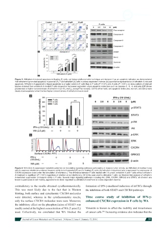
Figure 3: Withaferin A induced apoptosis in floating (F) cells. (a) Using cytofluorometric technique and Annexin V as an apoptotic indicator, we demonstrated
            that withaferin A promoted apoptosis in parental (P), F and adherent (A) cells in a dose-dependent manner; (b) quantitative representation of withaferin A-induced
            apoptosis. Withaferin A appeared to trigger apoptosis to a higher extent in F cells than in P cells (P < 0.01) or in A cells (P < 0.01); (c) immunoblots of total cell
            lysates obtained from withaferin A-treated F cells showed an increased expression of pro-apoptotic molecules such as caspase-3, -8, -9, and poly ADP-ribose
            polymerase at higher concentrations of withaferin A (5-10 μmol/L), except Fas receptor. On the other hand, anti-apoptotic molecules, survivin, and Bcl-2 were
            clearly down-regulated when the two higher concentrations of withaferin A were used


            Figure 4: Withaferin A suppressed metastatic potential via modulating signaling pathways participating in invasive tumor activity. (a) Withaferin A treatment was
            able to suppress interferon-γ-induced invasive ability in both parental (P) and to floating (F) cells; (b) withaferin A treatment also suppressed the expression of
            CXCR4 expression even under the stimulation of interferon-γ. The difference between F cells treated with 2.5 μmol/L withaferin A and F cells without withaferin
            A treatment is significant (P < 0.01) regardless of whether or not interferon-γ (10 U/mL) was used to stimulate F cells; (c) Western blot analysis of withaferin
            A-mediated suppression in invasive ability in F cells. Several major signaling pathways including Akt, ERK, CXCR4, GRK3/2 and STAT3, all of which are
            known to participate in cell mobility, appeared to be down-regulated by withaferin A treatment in a dose-dependent manner
            contradictory to the results obtained cytofluorometrically.   formation of IFN-γ-mediated induction of mCSCs through
            This was most likely  due to the fact that in  Western   the inhibition of both STAT3 and CXCR4 pathways.
            blotting, both surface and cytoplasmic CXCR4 molecules
            were  detected,  whereas  in  the  cytofluorometric  results,   Time  course  study  of  inhibition  of  IFN-γ-
            only the surface CXCR4 molecules were seen. Moreover,   enhanced CXCR4 expression in F cells by WA
            the inhibitory effect on the phosphorylation of STAT3 was
            readily noted at the highest concentration of WA (5 μmol/L)   Vimentin is known to affect the mobility and invasiveness
            used. Collectively, we concluded  that  WA blocked  the   of cancer cells.  Increasing evidence also indicates that the
                                                                          [26]

                         Journal of Cancer Metastasis and Treatment  ¦ Volume 2 ¦ Issue 1 ¦ January 15, 2016 ¦  35