
Page 43 - Read Online
P. 43
absence and presence of verapamil, the percentage of SP genes followed by P and A cells [Figure 1c], establishing F
cells in each group was calculated. Results of SP analysis cells as the major subpopulation containing the CSCs and
from 1 of 3 independent experiments showed that parental their CSC niche of the UP-LN1 cell line.
(P) UP-LN1 cells contained an intermediate of 2.93% SP
cells, A cells contained the least among the three groups at WA reduces SP and cell aggregates in UP-LN1 cells
1.07%, and F cells contained the highest of 4.20% [Figure
1a, upper frame]. The results show Hoechst 33342 dye We next sought to examine the potential CSC inhibitory
exclusion was verapamil-sensitive; they suggest that F cells effect of WA. Our cytofluorometric data demonstrated
contained the highest proportion of CSCs in UP-LN1, and that WA reduced the percentage of SP cells in UP-LN1
A cells contained the least. Quantitative results based on in a dose-dependent manner [Figure 2a]. The ability of
the 3 experiments reveal the statistical differences between WA to affect UP-LN1 viability was then tested on F and
F versus A and between F versus P cells in terms of the A cells. The viability of SP in A cells was affected least
percentage of SP cells as follows: F > A with P < 0.01, and among the three groups. WA preferentially targeted F cells
F > P with P < 0.05 [Figure 1a, lower frame]. To reinforce in a dose-dependent fashion [Figure 2b]. Since F cells
our SP data, an embryonic stem cell specific fluorescent dye spontaneously formed grape-like cell aggregates, they are a
CDy1 was used to stain UP-LN1 cells. The red fluorescent close representation of the so-called tumor spheres or CSCs
[22]
signal was strongly associated with F cells as compared to A reported. WA treatment also prevented the formation of
cells [arrowheads, Figure 1b]. Notably, red fluorescence was F-cell aggregates [Figure 2c]. At 10 µmol/L, WA reduced
significantly stronger in F cell aggregates [arrowheads, Figure F-cell aggregates by approximately 80%.
1b]. To add support to F cells identity as potential CSCs, we
examined the expression of stemness gene signatures such WA preferentially induces apoptosis in F cells
as Nanog, Oct4, Sox2, and c-Myc in the UP-LN1 cell line. F
cells exhibited the highest expression level of these stemness Since WA has been shown to induce apoptosis in cervical
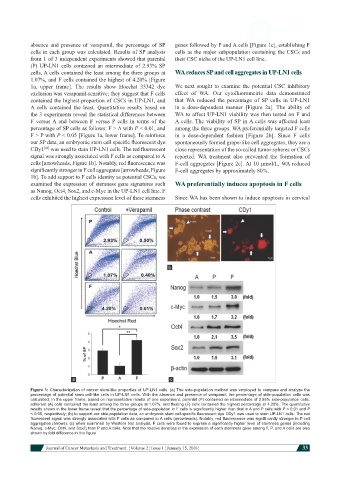
Figure 1: Characterization of cancer stem-like properties of UP-LN1 cells. (a) The side-population method was employed to compare and analyze the
percentage of potential stem cell-like cells in UP-LN1 cells. With the absence and presence of verapamil, the percentage of side-population cells was
calculated. In the upper frame, based on representative results of one experiment, parental (P) contained an intermediate of 2.93% side-population cells,
adherent (A) cells contained the least among the three groups at 1.07%, and floating (F) cells contained the highest percentage at 4.20%. The quantitative
results shown in the lower frame reveal that the percentage of side-population in F cells is significantly higher than that in A and P cells with P < 0.01 and P
< 0.05, respectively; (b) to support our side-population data, an embryonic stem cell-specific fluorescent dye CDy1 was used to stain UP-LN1 cells. The red
fluorescent signal was strongly associated with F cells as compared to A cells (arrowheads). Notably, red fluorescence was signifi cantly stronger in F-cell
aggregates (arrows); (c) when examined by Western blot analysis, F cells were found to express a significantly higher level of stemness genes (including
Nanog, c-Myc, Oct4, and Sox2) than P and A cells. Note that the relative densities in the expression of each stemness gene among F, P, and A cells are also
shown by fold difference in this figure
Journal of Cancer Metastasis and Treatment ¦ Volume 2 ¦ Issue 1 ¦ January 15, 2016 ¦ 33