
Page 29 - Read Online
P. 29
Frame et al. Investigating models for prostate cancer research
QPI label-free imaging technique. We used a panel of metastasis), LNCaP (lymph node metastasis) and
cell lines from a variety of sources, PNT2-C2 (normal), compared them to a primary culture [Figure 4A]. A
BPH-1 (benign), P4E6 (localized cancer), PC3 (bone 72-h time-lapse experiment was performed (images
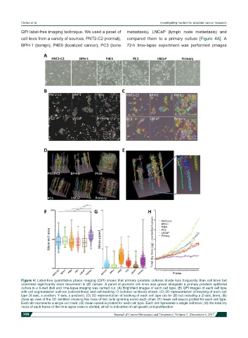
Figure 4: Label-free quantitative phase imaging (QPI) shows that primary prostate cultures divide less frequently than cell lines but
undertake significantly more movement in 2D culture. A panel of prostate cell lines was grown alongside a primary prostate epithelial
culture in a 6-well dish and time-lapse imaging was carried out. (A) Brightfield images of each cell type; (B) QPI images of each cell type
with cell segmentation outlines (colored lines) and cell tracking ID (colored numbers) shown; (C) 2D representation of tracking of each cell
type (X-axis, x position; Y-axis, y position); (D) 3D representation of tracking of each cell type (as for 2D but including a Z-axis, time); (E)
close up view of the 3D rendition showing the trace of two cells spinning round each other; (F) mean cell area is plotted for each cell type.
Each dot represents a single cell track; (G) mean speed is plotted for each cell type. Each dot represents a single cell track; (H) the total dry
mass of each frame of the time-lapse video is plotted, which is indicative of cell growth and proliferation
308 Journal of Cancer Metastasis and Treatment ¦ Volume 3 ¦ December 6, 2017