
Page 326 - Read Online
P. 326
Loria et al. CEUS in evaluation of vascularization of HCC
screening option. disc (CD) to be evaluated again. In no case were
complications manifested.
The development of second-generation ultrasound
contrast media and dedicated software has MSCT
improved the diagnostic capabilities of ultrasound The examinations used a CT multidetector scanner of
in the individualization and characterization of focal GE light-speed (16 and 64 canals). All examinations
hepatic lesions, [6-10] as it allows clinicians to study were done in basal conditions and after intravenous
intralesional vascular architecture in real time in all injection of approximately 90-120 mL of MdC, at
contrastographic phases. [11-15] a 4 mL/s speed. Smart prep was always used for
acquisition of the arterial phase.
Each contrastographic phase has its own specificity,
useful in diagnosis; in particular, in the arterial phase, Analysis of the images
it is fundamental to evaluate the pattern and the grade The vascularization of the single lesion, using both
of vascularization, while the portal and late phase are CEUS and CT, was classified as hyper-, iso- and
also useful for the correct diagnosis.
hypovascular in each one of the evaluated phases,
always in relation to the enhancement of the condition
METHODS of surrounding parenchyma.
Between January 2009 and May 2014, 67 patients
affected by hepatocarcinoma, who presented an Statistical analysis
overall of 92 nodules, were examined and enrolled in Fisher’s test was used to compare the results of
the study. There were 23 females and 44 males with CEUS with MSCT. Furthermore, the results of the
an average age of 68 years, of whom 62 presented vascularization comparing CEUS and MSCT were
a chronic liver disease, while 5 did not present evaluated in relation to the site and the size of
any hepatic symptoms. The diagnosis of HCC was the lesions. A P-value of < 0.05 was considered
established by confirming the presence of a lesion, statistically significant.
which assumed enhancement in the arterial phase
with wash-out in portal and late phases. The same RESULTS
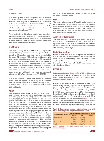
parameters were utilized in both contrast-enhanced In the arterial phase [Table 1], 75 of 92 nodules were
ultrasound (CEUS) and in multislice CT (MSCT).
hyperdense in MSCT; of these in using CEUS, 66
(88%) were hypervascular [Figure 1] and 9 (12%)
The three vascular phases were evaluated: arterial were isovascular [Figure 2].
(0-35 s from the injection of the MdC), portal (35-90 s)
and sinusoidal (from 90 s to approximately 6 min). Eleven of 92 nodules were isodense in MSCT; of
Increases in serum levels of alpha-fetoprotein were these using CEUS, 3 (27%) were hypervascular
also evaluated. [Figure 3], 8 (73%) were isovascular. Six of 92
nodules were hyperdense in MSCT, using CEUS,
CEUS 2 (33%) were hypervascular, 4 (67%) isovascular.
The examinations used GE LOGIQ 5 EXPERT Seventy-six of 92 nodules were localized at a depth
and ESAOTE MY LAB equipment with a specific not greater than 9 cm from the abdominal wall, 16 of
incorporated software designed to work at low 92 were localized at a greater depth [Table 2]. Of the
mechanical index, with 3.5 MHz convex transducers. 64 out of 76 nodules localized at a depth not greater
The contrast medium in all cases was the SonoVue than 9 cm that appeared hypervascular using MSCT,
(Bracco, Italy), consisting of micro bubbles of stabilized 61 (95%) appeared hypervascular using CEUS. Of the
phospholipids containing sulphur hexafluoride. This
was injected as a bolus in an antecubital vein, followed Table 1: Comparison between arterial phase seen with
by an injection of 10 mL of physiological solution. In MSCT and early vascular phase seen with CEUS
no case was a second injection of MdC given. Before Early vascular phase seen in CEUS
the intravenous injection, a basal echography of the Arterial phase in MSCT Hyper Iso Total
liver was done in order to evaluate the most suitable Hypervascular 66 (88%) 9 (12%) 75
ultrasound window to study the lesion. The identified Isovascular 3 (27%) 8 (73%) 11
lesion was studied “in real time” up to approximately Hypovascular 2 (33%) 4 (67%) 6
6 min from the injection and the enhancement was Total 71 (77%) 21 (23%) 92
always compared with the surrounding parenchyma. MSCT: multislice computed thomography; CEUS: contrast
All phases of the tests were registered on a compact enhanced ultrasound
Hepatoma Research ¦ Volume 2 ¦ November 22, 2016 317