
Page 22 - Read Online
P. 22
Fowler et al. Hepatoma Res 2020;6:19 I http://dx.doi.org/10.20517/2394-5079.2020.21 Page 7 of 10
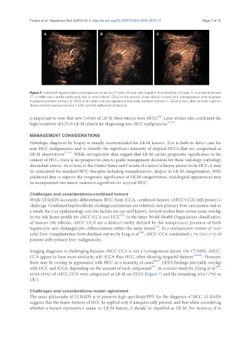
A B C
Figure 7. Combined hepatocellular-cholangiocarcinoma in a 77-year-old man with hepatitis-B related liver cirrhosis. A: contrast-enhanced
CT or MRI could not be performed due to renal failure. CEUS in the arterial phase shows a mass with homogeneous arterial phase
hyperenhancement (arrow); B: CEUS at 31 s after contrast injection shows early washout (arrow); C: CEUS at 60 s after contrast injection
shows marked washout (arrow). CEUS: contrast enhanced ultrasound
[38]
is important to note that 48% (39/82) of LR-M observations were HCCs . Later studies also confirmed the
high sensitivity of CEUS LR-M criteria for diagnosing non-HCC malignancies [39-41] .
MANAGEMENT CONSIDERATIONS
Histologic diagnosis by biopsy is usually recommended for LR-M lesions. This is both to direct care for
non-HCC malignancies and to identify the significant minority of atypical HCCs that are categorized as
LR-M observations [17,18] . While retrospective data suggest that LR-M carries prognostic significance in the
context of HCC, there is no prospective data to guide management decisions for these radiology-pathology
discordant lesions. As of now, in the United States and Canada, if a lesion is biopsy proven to be HCC, it may
be considered for standard HCC therapies including transplantation, despite its LR-M categorization. With
additional data to support the prognostic significance of LR-M categorization, radiological appearances may
be incorporated into future treatment algorithms for atypical HCC.
Challenges and considerations-combined tumors
While LI-RADS accurately differentiates HCC from iCCA, combined tumors (cHCC-CCA) still present a
challenge. Combined hepatocellular cholangiocarcinomas are relatively rare primary liver carcinomas and as
a result, the true epidemiology and risk factors are less well known. Several studies have shown some overlap
[42]
in the risk factor profile for cHCC-CCA and HCC . In the latest World Health Organization classification
of tumors 5th edition, cHCC-CCA are a distinct entity defined by the unequivocal presence of both
[43]
hepatocytic and cholangiocytic differentiation within the same tumor . In a retrospective review of 3103
[44]
adult liver transplantation from database entries by Jung et al. , cHCC-CCA constituted 2.7% (32/1173) of
patients with primary liver malignancies.
Imaging diagnosis is challenging because cHCC-CCA is not a homogeneous tumor. On CT/MRI, cHCC-
CCA appear to have more similarity with iCCA than HCC, often showing targetoid features [4,45,46] . However,
there may be overlap in appearance with HCC in a minority of cases [4,45] . CEUS findings inevitably overlap
[47]
[48]
with HCC and iCCA, depending on the amount of each component . In a recent study by Zheng et al. ,
20/24 (83%) of cHCC-CCA were categorized as LR-M on CEUS [Figure 7] and the remaining 4/24 (17%) as
LR-5.
Challenges and considerations-reader agreement
The main philosophy of LI-RADS is to preserve high specificity/PPV for the diagnosis of HCC. LI-RADS
suggests that the major features of HCC be applied only if unequivocally present, and that when considering
whether a feature represents a major vs. LR-M feature, it should be classified as LR-M. For instance, if in