
Page 18 - Read Online
P. 18
Fowler et al. Hepatoma Res 2020;6:19 I http://dx.doi.org/10.20517/2394-5079.2020.21 Page 3 of 10
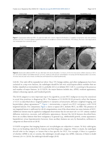
Figure 1. Gadoxetate enhanced MRI: 55 year-old male with chronic hepatitis B infection. A targetoid observation (65 mm) exhibits
features of LR-M: rim arterial phase hyperenhancement and peripheral washout (arrowheads). After biopsy, the lesion was diagnosed as
liver metastasis from colorectal cancer
Figure 2. Gadoxetate enhanced MRI: 65 year-old male with chronic hepatitis C cirrhosis. A 60-mm observation exhibits features of LR-5:
non rim arterial phase hyperenhancement (arrows), washout and capsule (arrowheads) indicating definite hepatocellular carcinoma.
Ancillary features seen are a mosaic architecture and hepatobiliary phase hypointensity
(LR-M). This task will be expanded on below. Once TIV, benign entities, and other malignancies have been
excluded in a step-wise fashion, the radiologist should be left with solid hepatocellular nodules that are
further classified as intermediate (LR-3), probable (LR-4) or definite HCC (LR-5), according to the presence
and number of major features. In LI-RADS, the major features include size, APHE, washout appearance,
delayed enhancing capsule, and threshold growth.
The LR-M category is a very important step in the algorithm, as non-HCC malignancies must be considered
to avoid false positives in diagnosing LR-5. The features of LI-RADS LR-M primarily reflect the features
of iCCA as described above: targetoid patterns in dynamic enhancement, diffusion weighted imaging, and
[15]
hepatobiliary phase appearances . Figure 1 demonstrates a typical non-HCC malignancy with LR-M
targetoid features. For comparison, Figure 2 shows a typical LR-5, definite HCC, on MRI. The presence of
any targetoid feature is sufficient for LR-M categorization, even if the observation has some features of HCC.
Additional features may be applied in malignant observations that do not meet LR-5 or TIV criteria: marked
diffusion restriction, necrosis, and infiltrative appearances. Beyond the LR-M features described here,
there are ancillary features that favor malignancy in general (e.g., subthreshold growth, corona appearance,
hepatobiliary phase hypointensity); however, these ancillary features are not, by themselves, sufficient to
categorize an observation as LR-M.
LI-RADS recognizes that imaging features and morphologies are subjective in nature. To help radiologists,
there are tie breaking rules both for features and final diagnostic categories. When in doubt, the radiologist
should refer to the category or feature that is less specific for HCC. For example, if there is a question
of whether APHE is rim or nonrim, the radiologist should assign rim APHE. Likewise, if there is doubt
between LR-5 vs. LR-M, the radiologist should assign LR-M.