
Page 21 - Read Online
P. 21
Page 6 of 10 Fowler et al. Hepatoma Res 2020;6:19 I http://dx.doi.org/10.20517/2394-5079.2020.21
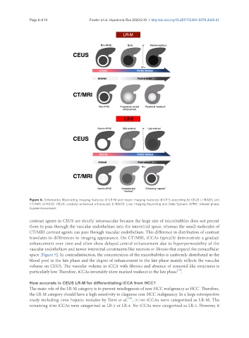
Figure 6. Schematics illustrating imaging features of LR-M and major imaging features of LR-5 according to CEUS LI-RADS and
CT/MRI LI-RADS. CEUS: contrast enhanced ultrasound; LI-RADS: Liver Imaging Reporting and Data System; APHE: arterial phase
hyperenhancement
contrast agents in CEUS are strictly intravascular because the large size of microbubbles does not permit
them to pass through the vascular endothelium into the interstitial space, whereas the small molecules of
CT/MRI contrast agents can pass through vascular endothelium. This difference in distribution of contrast
translates to differences in imaging appearance. On CT/MRI, iCCAs typically demonstrate a gradual
enhancement over time and often show delayed central enhancement due to hyperpermeability of the
vascular endothelium and tumor interstitial constituents like necrosis or fibrosis that expand the extracellular
space [Figure 5]. In contradistinction, the concentration of the microbubbles is uniformly distributed in the
blood pool in the late phase and the degree of enhancement in the late phase mainly reflects the vascular
volume on CEUS. The vascular volume in iCCA with fibrosis and absence of sinusoid-like structures is
[37]
particularly low. Therefore, iCCAs invariably show marked washout in the late phase .
How accurate is CEUS LR-M for differentiating iCCA from HCC?
The main role of the LR-M category is to prevent misdiagnosis of non-HCC malignancy as HCC. Therefore,
the LR-M category should have a high sensitivity to diagnose non-HCC malignancy. In a large retrospective
[38]
study including 1006 hepatic nodules by Terzi et al. , 31/40 iCCAs were categorized as LR-M. The
remaining 9/40 iCCAs were categorized as LR-3 or LR-4. No iCCAs were categorized as LR-5. However, it