
Page 119 - Read Online
P. 119
Page 6 of 14 Santillan Hepatoma Res 2020;6:63 I http://dx.doi.org/10.20517/2394-5079.2020.60
A B
C D
E F
G H
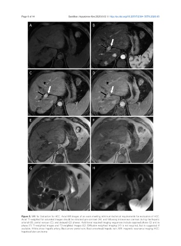
Figure 3. MRI for Evaluation for HCC. Axial MR images of an exam meeting minimum technical requirements for evaluation of HCC.
Axial T1-weighted fat saturated images should be obtained pre-contrast (A), and following intravenous contrast during the hepatic
arterial (B), portal venous (C), and delayed (D) phases. Additional required imaging sequences include opposed phase (E) and in
phase (F) T1-weighted images and T2-weighted images (G). Diffusion weighted imaging (H) is not required, but is suggested if
available. White arrow: hepatic artery; Black arrow: portal vein; Black arrowhead: hepatic vein. MRI: magnetic resonance imaging; HCC:
hepatocellular carcinoma