
Page 116 - Read Online
P. 116
Santillan Hepatoma Res 2020;6:63 I http://dx.doi.org/10.20517/2394-5079.2020.60 Page 3 of 14
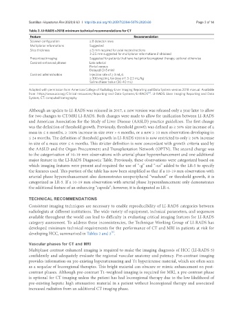
Table 2. LI-RADS v2018 minimum technical recommendations for CT
Feature Recommendation
Scanner configuration ≥ 8 detector rows
Multiplanar reformations Suggested
Slice thickness ≤ 5 mm required for axial reconstructions
3-2.5 mm suggested for multiplanar reformations if obtained
Precontrast imaging Suggested for patients that have had prior locoregional therapy, optional otherwise
Contrast-enhanced phases Late arterial
Portal venous
Delayed (2-5 min)
Contrast administration Injection rate of ≥ 3 mL/s
≥ 300 mgI/mL for dose of 1.5-2.5 mL/kg
Saline chaser bolus (30-40 mL)
Adapted with permission from American College of Radiology Liver Imaging Reporting and Data System version 2018 manual. Available
[4]
from: https//www.acr.org/Clinical-resources/Reporting-and-Data-Systems/LI-RADS . LI-RADS: Liver Imaging Reporting and Data
System; CT: computed tomography
Although an update to LI-RADS was released in 2017, a new version was released only a year later to allow
for two changes to CT/MRI LI-RADS. Both changes were made to allow for unification between LI-RADS
and American Association for the Study of Liver Disease (AASLD) practice guidelines. The first change
was the definition of threshold growth. Previously, threshold growth was defined as ≥ 50% size increase of a
mass in ≤ 6 months, ≥ 100% increase in size over > 6 months, or a new ≥ 10 mm observation developing in
≤ 24 months. The definition of threshold growth in LI-RADS v2018 is now restricted to only ≥ 50% increase
in size of a mass over ≤ 6 months. This stricter definition is now concordant with growth criteria used by
the AASLD and the Organ Procurement and Transplantation Network (OPTN). The second change was
to the categorization of 10-19 mm observations with arterial phase hyperenhancement and one additional
major feature in the LI-RADS Diagnostic Table. Previously, these observations were categorized based on
which imaging features were present and required the use of “-g” and “-us” added to the LR-5 to specify
the features used. This portion of the table has now been simplified so that if a 10-19 mm observation with
arterial phase hyperenhancement also demonstrates nonperipheral “washout” or threshold growth, it is
categorized as LR-5. If a 10-19 mm observation with arterial phase hyperenhancement only demonstrates
the additional feature of an enhancing “capsule”; however, it is designated as LR-4.
TECHNICAL RECOMMENDATIONS
Consistent imaging techniques are necessary to enable reproducibility of LI-RADS categories between
radiologists at different institutions. The wide variety of equipment, technical parameters, and sequences
available throughout the world can lead to difficulty in evaluating critical imaging features for LI-RADS
category assessment. To address these inconsistencies, the Technique Working Group of LI-RADS has
developed minimum technical requirements for the performance of CT and MRI in patients at risk for
[5]
developing HCC, summarized in Tables 2 and 3 .
Vascular phases for CT and MRI
Multiphase contrast enhanced imaging is required to make the imaging diagnosis of HCC (LI-RADS 5)
confidently and adequately evaluate the regional vascular anatomy and patency. Pre-contrast imaging
provides information on pre-existing hyperattenuating and T1 hyperintense material, which are often seen
as a sequelae of locoregional therapies. This bright material can obscure or mimic enhancement on post-
contrast phases. Although pre-contrast T1-weighted imaging is required for MRI, a pre-contrast phase
is optional for CT imaging unless the patient has had locoregional therapy due to the low likelihood of
pre-existing hepatic high attenuation material in a patient without locoregional therapy and associated
increased radiation from an additional CT imaging phase.