
Page 117 - Read Online
P. 117
Page 4 of 14 Santillan Hepatoma Res 2020;6:63 I http://dx.doi.org/10.20517/2394-5079.2020.60
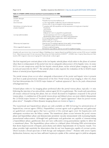
Table 3. LI-RADS v2018 minimum technical recommendations for MRI
Feature Recommendation
Scanner strength 1.5 or 3 Tesla
Coil type Phased array multichannel torso coil
Slice thickness ≤ 5 mm required for multiphase contrast-enhanced sequences
≤ 8 mm with ≤ 2 mm slice gap for other sequences
Multiphase contrast-enhanced T1 weighted with 3D acquisition and fat suppression strongly recommended
phases (required) Precontrast
Late arterial
Portal venous
Delayed (2-5 min) if using ECA or gadobenate
Contrast administration Transitional phase (2-5 min) if using gadoxetate
Hepatobiliary phase (approx 20 min) if using gadoxetate
ECA or gadobenate or gadoxetate, weight adjusted dose
Injection rate of 1-2 mL/s
Saline chaser bolus (30-40 mL)
Other required sequences Unenhanced T1-weighted opposed-phase and in-phase imaging
T2-weighted imaging (fat suppression optional)
Other suggested sequences Diffusion weighted imaging
Multiplanar acquisitions, such as coronal T2-weighted imaging
Adapted with permission from American College of Radiology Liver Imaging Reporting and Data System version 2018 manual. Available
[4]
from: https//www.acr.org/Clinical-resources/Reporting-and-Data-Systems/LI-RADS . LI-RADS: Liver Imaging Reporting and Data
System; MRI: magnetic resonance imaging; ECA: extracellular contrast agent
The first required post-contrast phase is the late hepatic arterial phase which refers to the phase of contrast
when there is enhancement of the portal vein but no antegrade enhancement of the hepatic veins. As some
HCCs are not conspicuous until the late hepatic arterial phase, earlier arterial phase imaging can result
in reduced sensitivity for HCC . The arterial phase is also required for evaluation of the LI-RADS major
[6]
feature of arterial phase hyperenhancement.
The portal venous phase occurs when antegrade enhancement of the portal and hepatic veins is present
and there is peak parenchymal enhancement of the liver. Portal venous phase imaging is often the phase
that best demonstrates the LI-RADS major feature of “washout appearance” due to the peak enhancement
of background liver.
Delayed phase refers to the imaging phase performed after the portal venous phase, typically 3-5 min
following the injection of an extracellular contrast agent (ECA) or gadobenate. The vessels and parenchyma
all remain enhanced during this phase, but are overall decreased in brightness compared to the portal
venous phase. A combination of the portal venous phase and delayed phase can more reliably demonstrate
the LI-RADS major features of “washout appearance” and “capsule appearance” than the portal venous
[7]
phase alone . Examples of these dynamic imaging phases are shown in Figure 2.
The transitional and hepatobiliary phases are only available on MRI following the administration of
hepatobiliary contrast agents (HBAs). Hepatobiliary contrast agents such as gadobenate and gadoxetate
are gadolinium contrast agents that have a portion of the contrast agent enter the hepatocytes with
subsequent excretion into the biliary system. The transitional phase occurs between the portal venous
phase and hepatobiliary phase and demonstrates persistent vascular enhancement with increasing hepatic
parenchymal enhancement. Although both gadobenate and gadoxetate are capable of demonstrating
a hepatobiliary phase, gadobenate does not have sufficiently strong parenchymal update to allow for
a transitional phase. The hepatobiliary phase refers to a delayed phase following HBA administration
that shows hyperintensity of hepatic parenchyma and relative hypointensity of the hepatic vessels. This
phase provides high sensitivity for observations that do not have hepatocytes capable of contrast uptake,
[7]
which includes a broad spectrum of both benign and malignant entities . Patients with severe hepatic
dysfunction, however, may not have sufficient hepatocyte uptake of the contrast agent to yield an adequate