
Page 122 - Read Online
P. 122
Santillan Hepatoma Res 2020;6:63 I http://dx.doi.org/10.20517/2394-5079.2020.60 Page 9 of 14
A B
C D
E F
G H
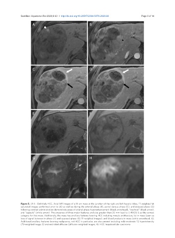
Figure 5. LR-5 - Definitely HCC. Axial MR images of a 14 cm mass at the junction of the right and left hepatic lobes. T1-weighted fat
saturated images performed prior to (A) as well as during the arterial phase (B), portal venous phase (C), and delayed phase (D)
following contrast administration demonstrate areas of arterial phase hyperenhancement (black arrowhead), “washout” (black arrow),
and “capsule” (white arrow). The presence of three major features and size greater than 20 mm lead to LI-RADS 5 as the correct
category for this mass. Additionally, the mass has ancillary features favoring HCC including mosaic architecture, fat in mass (seen as
loss of signal between in-phase (F) and opposed-phase (E) T1-weighted images), and blood products in mass (white arrowhead, A).
Additional ancillary features favoring malignancy, not HCC in particular, are also present including mild-moderate T2 hyperintensity
(T2-weighted image, G) and restricted diffusion (diffusion weighted images, H). HCC: hepatocellular carcinoma