
Page 124 - Read Online
P. 124
Santillan Hepatoma Res 2020;6:63 I http://dx.doi.org/10.20517/2394-5079.2020.60 Page 11 of 14
Table 4. LI-RADS v2018 ancillary imaging features
Features favoring malignancy, not HCC in particular Features favoring HCC in particular Features favoring benignity
US visibility as discrete nodule Nonenhancing “capsule” Size stability ≥ 2 years
Subthreshold growth Nodule-in-nodule architecture Size reduction
Corona enhancement Mosaic architecture Parallels blood pool enhancement
Fat sparing in solid mass Fat in mass, more than adjacent liver Undistorted vessels
Restricted diffusion Blood products in mass Iron in mass, more than liver
Mild-moderate T2 hyperintensity Marked T2 hyperintensity
Iron sparing in solid mass Hepatobiliary phase isointensity
Transitional phase hypointensity
Hepatobiliary phase hypointensity
RADS: Liver Imaging Reporting and Data System; HCC: hepatocellular carcinoma
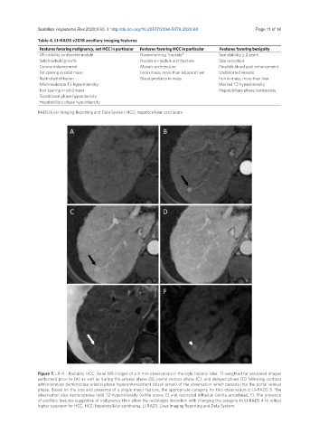
A B
C D
E F
Figure 7. LR-4 - Probably HCC. Axial MR images of a 9 mm observation in the right hepatic lobe. T1-weighted fat saturated images
performed prior to (A) as well as during the arterial phase (B), portal venous phase (C), and delayed phase (D) following contrast
administration demonstrate arterial phase hyperenhancement (black arrow) of the observation which persists into the portal venous
phase. Based on the size and presence of a single major feature, the appropriate category for this observation is LI-RADS 3. The
observation also demonstrates mild T2-hyperintensity (white arrow, E) and restricted diffusion (white arrowhead, F). The presence
of ancillary features suggestive of malignancy then allow the radiologist discretion with changing the category to LI-RADS 4 to reflect
higher suspicion for HCC. HCC: hepatocellular carcinoma; LI-RADS: Liver Imaging Reporting and Data System