
Page 125 - Read Online
P. 125
Page 12 of 14 Santillan Hepatoma Res 2020;6:63 I http://dx.doi.org/10.20517/2394-5079.2020.60
A B
C D
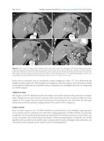
Figure 8. LR-TIV. Axial CT images from the hepatic arterial (A), portal venous (B), and delayed (C) phases following intravenous
contrast administration and coronal image from the hepatic arterial phase (D). Enhancing soft tissue is present in the right and main
portal veins (white arrow). Due to an associated parenchymal mass (black arrow) with arterial phase hyperenhancement (not shown)
and “washout”, the most appropriate category for this finding is LR-TIV, likely due to HCC. CT: computed tomography; LI-RADS: Liver
Imaging Reporting and Data System; HCC: hepatocellular carcinoma; LR-TIV: LI-RADS Tumor in Vein
[8]
lesions such as metastases from an extrahepatic primary malignancy [Figure 7] . If an observation has
multiple ancillary features for both benignity and malignancy, then the category of the observation should
not be adjusted. Finally, the use of ancillary features is optional at the radiologist’s discretion for designating
a LI-RADS category.
TUMOR IN VEIN
The category of LR-TIV should be used for the evidence of vascular invasion of the portal veins or hepatic
veins. Unequivocal vascular invasion is present when enhancing soft tissue is present within the vessel
[Figure 8]. This category does not require the presence of a parenchymal mass. If possible, the radiologist
should indicate the most probably etiology between HCC and non-HCC malignancy.
CONCLUSION
Since its initial release in 2011, CT/MR LI-RADS has evolved based on user feedback, ongoing expert
review, and the need for unification with other HCC imaging algorithms. CT/MR LI-RADS v2018 provides
an algorithm for the standardized reporting and interpretation of findings in patients at risk for HCC, and
is now concordant with AASLD practice guidelines. With increasing adoption worldwide, the CT/MR
algorithm and associated lexicon enable clearer communication between radiologists, other physicians, and
researchers to better provide care for patients at risk for developing HCC.