
Page 118 - Read Online
P. 118
Santillan Hepatoma Res 2020;6:63 I http://dx.doi.org/10.20517/2394-5079.2020.60 Page 5 of 14
A B
C D
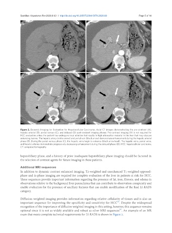
Figure 2. Dynamic Imaging for Evaluation for Hepatocellular Carcinoma. Axial CT images demonstrating the pre-contrast (A),
hepatic arterial (B), portal venous (C), and delayed (D) post-contrast imaging phases. Pre-contrast imaging (A) is not required for
HCC evaluation unless the patient has undergone local ablation that results in high attenuation material in the liver that may obscure
enhancing lesions. The hepatic artery (white arrow) and portal vein (black arrow) demonstrate enhancement during the hepatic arterial
phase (B). During the portal venous phase (C), the hepatic veins begin to enhance (black arrowhead). The hepatic veins, portal veins,
and hepatic arteries demonstrate progressively decreasing enhancement during the delayed phase (D). HCC: hepatocellular carcinoma;
CT: computed tomography
hepatobiliary phase, and a history of prior inadequate hepatobiliary phase imaging should be factored in
the selection of contrast agents for future imaging in these patients.
Additional MRI sequences
In addition to dynamic contrast enhanced imaging, T2-weighted and unenhanced T1-weighted opposed-
phase and in-phase imaging are required for complete evaluation of the liver in patients at risk for HCC.
These sequences provide important information regarding the presence of fat, iron, fibrosis, and edema in
observations relative to the background liver parenchyma that can contribute to observation conspicuity and
enable evaluation for the presence of ancillary features that can enable modification of the final LI-RADS
category.
Diffusion weighted imaging provides information regarding relative cellularity of tissues and is also an
important sequence for improving the specificity and sensitivity for HCC . Despite the widespread
[8]
recognition of the importance of diffusion weighted imaging in this setting, however, this sequence remains
[5]
optional since it is not as widely available and robust as other MRI sequences . An example of an MR
exam that meets complete technical requirements for LI-RADS is shown in Figure 3.