
Page 115 - Read Online
P. 115
Page 2 of 14 Santillan Hepatoma Res 2020;6:63 I http://dx.doi.org/10.20517/2394-5079.2020.60
Table 1. LI-RADS v2018 diagnostic categories
Category Definition
LR-1 Definitely benign
LR-2 Probably benign
LR-3 Intermediate probability of malignancy
LR-4 Probably HCC
LR-5 Definitely HCC
LR-M Probably or definitely malignancy but not HCC specific
LR-TIV Definite tumor in vein
LR-NC Cannot be categorized due to image degradation or omission
LI-RADS: Liver Imaging Reporting and Data System; HCC: hepatocellular carcinoma; LR-TIV: LI-RADS Tumor in Vein
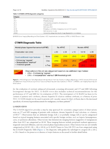
Figure 1. LI-RADS Diagnostic Table. Reproduced with permission from the American College of Radiology LI-RADS version 2018 manual.
[4]
Available from: https//www.acr.org/Clinical-resources/Reporting-and-Data-Systems/LI-RADS . LI-RADS: Liver Imaging Reporting and
Data System
for the evaluation of contrast-enhanced ultrasound, screening ultrasound, and CT and MRI following
locoregional therapy for HCC. LI-RADS v2018 also includes technical recommendations for the
performance of CT and MRI for the evaluation of HCC. The development of LI-RADS has been in the
context of patients with cirrhosis, chronic hepatitis B infection without cirrhosis, or a history of prior
HCC. LI-RADS should not be used in patients with vascular causes for their cirrhosis due to the decreased
[3]
specificity of arterial hyperenhancement for malignancy in these patients .
LI-RADS V2018
The LI-RADS algorithm provides a step by step approach for consistent categorization of observations
seen on CT and MR imaging of patients with cirrhosis, chronic hepatitis B viral infection, or a history
[4]
of HCC . Observations that are definitely benign (LR-1) or probably benign (LR-2) can be categorized
based on typical imaging features associated with specific benign entities, such as hepatic hemangiomas,
cysts, or perfusion alterations. Observations that demonstrate imaging features suggestive of malignancies
other than HCC are categorized as LR-M. These imaging features include peripheral washout appearance,
targetoid restricted diffusion, and rim arterial phase hyperenhancement. Observations that are not initially
categorized as LR-NC, LR-1, LR-2, LR-M, or LI-RADS Tumor in Vein (LR-TIV) are then categorized using
the LI-RADS Diagnostic Table [Figure 1]. The diagnostic table guides the radiologist to category LR-3, LR-4,
or LR-5 based on the presence or absence of major features as discussed in further detail below.