
Page 176 - Read Online
P. 176
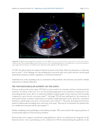
Alberts et al. Vessel Plus 2023;7:34  https://dx.doi.org/10.20517/2574-1209.2023.37   Page 15 of 24


                Figure 10. Modified mid-esophageal five-chamber view (0) with a focus on the right atrium. A large aortic root abscess presents
                (yellow arrows) with a fistula (white arrows) from the aortic root to the dilated right atrium. Continuous flow in systole and diastole is
                seen using color flow Doppler. LA: Left atrium; LV: left ventricle; RA: right atrium; RV: right ventricle.

               On TEE, the infected graft may appear thickened and para-aortic fluid collections, hematoma, or abscesses
               may be seen . Other findings on echocardiography associated with aortic graft infection include graft
                          [97]
               anastomotic disruption, fistulae, vegetations, or aneurysm formation .
                                                                         [95]
               Graft infections of the ascending aorta, in combination with prosthetic valve infection, may lead to valvular
               dysfunction or valvular dehiscence.


               TEE FOR ENDOVASCULAR SURGERY ON THE AORTA
               Thoracic endovascular aortic repair (TEVAR) has been used for two decades and has revolutionized the
               treatment for disease of the aorta. It is now the preferred approach for the treatment of aneurysms of the
               descending thoracic aorta, due to its reduced morbidity, hospital length of stay, and short-term mortality
               compared to open thoracic aneurysm repair . Although TEVAR was developed for the treatment of
                                                       [98]
               aneurysmal disease, treatment indications now include complicated type B aortic dissection, intramural
               hematoma, penetrating aortic ulcer, and traumatic aortic lesions [98,99] . Recently, developments have been
               made for endovascular ascending aortic and aortic arch repair. This may be an alternative for patients who
               are considered at increased risk for open surgery .
                                                        [100]

               Besides visualizing aortic pathologies as described previously, TEE can be used for the surgical guidance of
               several specific aspects of endovascular procedures:

               Endovascular aortic surgery is performed using guidewires, which are usually placed retrograde via the
               femoral arteries. Correct positioning can be confirmed by TEE by demonstrating the guidewire in the