
Page 172 - Read Online
P. 172
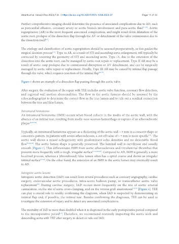
Alberts et al. Vessel Plus 2023;7:34  https://dx.doi.org/10.20517/2574-1209.2023.37   Page 11 of 24

               Further comprehensive imaging should determine the presence of associated complications due to AD, such
               as pericardial effusion, coronary artery or aortic branch involvement and para-aortic fluid [11,72] . Aortic
               regurgitation (AR) is the most frequent associated complication, and might result from dilatation of the
               aortic root, prolapse of the dissection flap through the AV or detachment of the valve commissures due to
               the dissection itself .
                               [66]
               The etiology and classification of aortic regurgitation should be assessed preoperatively, as this guides the
               surgical decision process . Type 1a AR, as a result of STJ and ascending aorta enlargement, will typically be
                                    [73]
               corrected by restoring the geometry of STJ and ascending aorta. Type 1b, due to the extension of the
               dissection into the aortic root, can be managed by aortic root repair or replacement. Type II AR may be a
               result of aortic cusp prolapse due to commissural disruption or AV detachment, and can be surgically
               managed by aortic valve repair or replacement. Finally, Type III AR may be caused by intimal flap passage
               through the valve, which requires resection of the intimal flap [64,73] .


               Figure 5 shows an example of a dissection flap passing through the aortic valve.

               After surgery, the evaluation of the repair with TEE includes aortic valve function, coronary flow detection,
               and regional wall motion abnormalities. The flow in the aortic lumens should be assessed by the
               echocardiographist to determine the correct flow in the true lumen and to rule out a residual connection
               between the true and false lumen.

               Intramural hematoma
               An intramural hematoma (IMH) occurs when blood collects in the media of the aortic wall, with the
               absence of an intimal tear, resulting from media vasa vasorum hemorrhage or rupture of an atherosclerotic
               plaque [18,68,74] .


               Typically, an intramural hematoma appears as a thickening of the aortic wall > 5 mm in a crescent-shape or
               concentric pattern. In patients with severe atherosclerosis, a cut-off value of > 7 mm is more specific . The
                                                                                                    [17]
               aortic wall shows a mixed echogenicity with predominant echo densities and no detectable blood
               flow [18,68,74] . The aortic lumen shape is generally preserved. The luminal wall is curvilinear and usually
               smooth [Figure 7]. This differentiates IMH from aortic atherosclerosis and intraluminal thrombus that
               presents more frequently with a rough, irregular surface [17,18,20,74] . Compared to AD, IMH is generally a more
               localized process, whereas a (thrombosed) false lumen often has a spiral course and shows an irregular
               intimal surface [17,74] . On the other hand, the extension of an IMH to the aortic lumen may eventually result
               in AD.


               Iatrogenic aortic lesions
               Iatrogenic aortic dissection (IAD) can result from several procedures such as coronary angiography, cardiac
               surgery, endovascular aortic procedures, intra-aortic balloon pump, or transcatheter aortic valve
               replacement . During cardiac surgery, IAD occurs most frequently on the site of aortic arterial
                         [75]
               cannulation, on the site of aorta cross-clamping, and on the venous graft anastomosis [18,75]  [Figure 8]. TEE
               can play a crucial role in readily confirming the diagnosis, when IAD is suspected by demonstrating an
               intimal flap and, if possible, the intimal tear. Besides confirming the diagnoses, TEE can be used to
               investigate the extension of injury and to detect any associated complications.


               The mortality of IAD is more than doubled when it is diagnosed in the early postoperative period compared
                                         [76]
               to the intraoperative period . Therefore, we recommend routinely inspecting the aortic arch and
               descending aorta with TEE after surgery to detect or rule out IAD.