
Page 169 - Read Online
P. 169
Page 8 of 24 Alberts et al. Vessel Plus 2023;7:34 https://dx.doi.org/10.20517/2574-1209.2023.37
Figure 3. Descending aorta short axis view. An enlarged descending aorta can be seen, with the Frozen Elephant Trunk clearly visible in
the lumen. Right image with color flow Doppler with adequate flow in the Frozen Elephant Trunk. AoDs: Descending aorta; FET: frozen
elephant trunk.
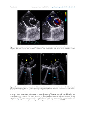
Figure 4. Simultaneous multiplane image with (A) Mid-esophageal descending aorta short axis view (0) and (B) Mid-esophageal
descending aorta long axis view (90). Yellow arrows: Severe atherosclerosis (grade IV) with intimal thickening > 10 mm and irregular
border. Blue arrows: Multiple reverberation artefacts from the calcified aortic (comet-tails).
Preoperatively, it is important to document the size and location of the aneurysm with TEE, although it can
be challenging to measure the exact diameter of the dilated aorta due to off-axis imaging. Aortic
regurgitation (AR) is a common secondary complication of aortic root and ascending aorta dilatation and
can be severe . When present, the severity and etiology of AR should be assessed with TEE.
[64]