
Page 173 - Read Online
P. 173
Page 12 of 24 Alberts et al. Vessel Plus 2023;7:34 https://dx.doi.org/10.20517/2574-1209.2023.37
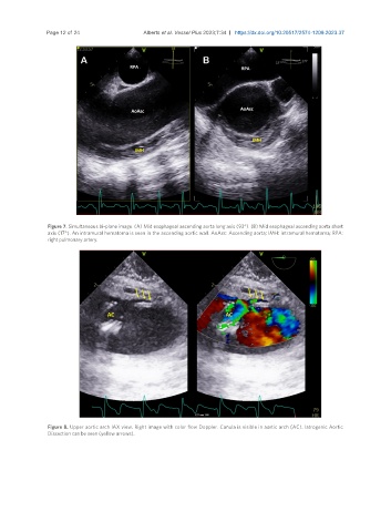
Figure 7. Simultaneous bi-plane image. (A) Mid esophageal ascending aorta long axis (93°). (B) Mid esophageal ascending aorta short
axis (17°). An intramural hematoma is seen in the ascending aortic wall. AoAsc: Ascending aorta; IMH: intramural hematoma; RPA:
right pulmonary artery.
Figure 8. Upper aortic arch lAX view. Right image with color flow Doppler. Canula is visible in aortic arch (AC). Iatrogenic Aortic
Dissection can be seen (yellow arrows).