
Page 175 - Read Online
P. 175
Page 14 of 24 Alberts et al. Vessel Plus 2023;7:34 https://dx.doi.org/10.20517/2574-1209.2023.37
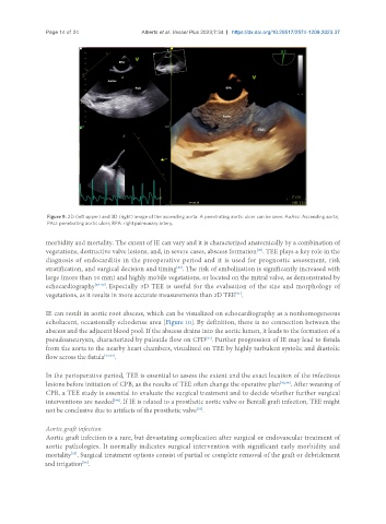
Figure 9. 2D (left upper) and 3D (right) image of the ascending aorta. A penetrating aortic ulcer can be seen. AoAsc: Ascending aorta;
PAU: penetrating aortic ulcer; RPA: right pulmonary artery.
morbidity and mortality. The extent of IE can vary and it is characterized anatomically by a combination of
[85]
vegetations, destructive valve lesions, and, in severe cases, abscess formation . TEE plays a key role in the
diagnosis of endocarditis in the preoperative period and it is used for prognostic assessment, risk
stratification, and surgical decision and timing . The risk of embolization is significantly increased with
[86]
large (more than 10 mm) and highly mobile vegetations, or located on the mitral valve, as demonstrated by
echocardiography [87-90] . Especially 3D TEE is useful for the evaluation of the size and morphology of
vegetations, as it results in more accurate measurements than 2D TEE .
[91]
IE can result in aortic root abscess, which can be visualized on echocardiography as a nonhomogeneous
echolucent, occasionally echodense area [Figure 10]. By definition, there is no connection between the
abscess and the adjacent blood pool. If the abscess drains into the aortic lumen, it leads to the formation of a
pseudoaneurysm, characterized by pulsatile flow on CFD . Further progression of IE may lead to fistula
[92]
from the aorta to the nearby heart chambers, visualized on TEE by highly turbulent systolic and diastolic
flow across the fistula [92,93] .
In the perioperative period, TEE is essential to assess the extent and the exact location of the infectious
lesions before initiation of CPB, as the results of TEE often change the operative plan [92,94] . After weaning of
CPB, a TEE study is essential to evaluate the surgical treatment and to decide whether further surgical
interventions are needed . If IE is related to a prosthetic aortic valve or Bentall graft infection, TEE might
[94]
[75]
not be conclusive due to artifacts of the prosthetic valve .
Aortic graft infection
Aortic graft infection is a rare, but devastating complication after surgical or endovascular treatment of
aortic pathologies. It normally indicates surgical intervention with significant early morbidity and
mortality . Surgical treatment options consist of partial or complete removal of the graft or debridement
[95]
and irrigation .
[96]