
Page 179 - Read Online
P. 179
Page 18 of 24 Alberts et al. Vessel Plus 2023;7:34 https://dx.doi.org/10.20517/2574-1209.2023.37
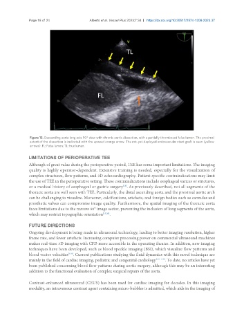
Figure 13. Descending aorta long axis 90° view with chronic aortic dissection, with a partially thrombosed false lumen. The proximal
extent of the dissection is indicated with the upward orange arrow. The not-yet-deployed endovascular stent graft is seen (yellow
arrows). FL: False lumen; TL: true lumen.
LIMITATIONS OF PERIOPERATIVE TEE
Although of great value during the perioperative period, TEE has some important limitations. The imaging
quality is highly operator-dependent. Extensive training is needed, especially for the visualization of
complex structures, flow patterns, and 3D echocardiography. Patient-specific contraindications may limit
the use of TEE in the perioperative setting. These contraindications include esophageal varices or strictures,
[13]
or a medical history of esophageal or gastric surgery . As previously described, not all segments of the
thoracic aorta are well seen with TEE. Particularly, the distal ascending aorta and the proximal aortic arch
can be challenging to visualize. Moreover, calcifications, artefacts, and foreign bodies such as cannulas and
prosthetic valves can compromise image quality. Furthermore, the spatial imaging of the thoracic aorta
faces limitations due to the narrow 90° image sector, preventing the inclusion of long segments of the aorta,
which may restrict topographic orientation [17,20] .
FUTURE DIRECTIONS
Ongoing development is being made in ultrasound technology, leading to better imaging resolution, higher
frame rate, and fewer artefacts. Increasing computer processing power on commercial ultrasound machines
makes real-time 3D imaging with CFD more accessible in the operating theater. In addition, new imaging
techniques have been developed, such as blood speckle imaging (BSI), which visualize flow patterns and
[110]
blood vector velocities . Current publications studying the fluid dynamics with this novel technique are
mainly in the field of cardiac imaging, pediatric and congenital cardiology [111-113] . To date, no articles have yet
been published concerning blood flow patterns during aortic surgery, although this may be an interesting
addition to the functional evaluation of complex surgical repairs of the aorta.
Contrast-enhanced ultrasound (CEUS) has been used for cardiac imaging for decades. In this imaging
modality, an intravenous contrast agent containing micro-bubbles is admitted, which aids in the imaging of