
Page 178 - Read Online
P. 178
Alberts et al. Vessel Plus 2023;7:34 https://dx.doi.org/10.20517/2574-1209.2023.37 Page 17 of 24
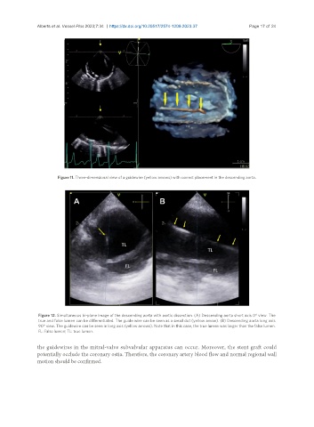
Figure 11. Three-dimensional view of a guidewire (yellow arrows) with correct placement in the descending aorta.
Figure 12. Simultaneous bi-plane image of the descending aorta with aortic dissection. (A) Descending aorta short axis 0° view. The
true and false lumen can be differentiated. The guide wire can be seen as a small dot (yellow arrow). (B) Descending aorta long axis
90° view. The guidewire can be seen in long axis (yellow arrows). Note that in this case, the true lumen was larger than the false lumen.
FL: False lumen; TL: true lumen.
the guidewires in the mitral-valve subvalvular apparatus can occur. Moreover, the stent graft could
potentially occlude the coronary ostia. Therefore, the coronary artery blood flow and normal regional wall
motion should be confirmed.