
Page 171 - Read Online
P. 171
Page 10 of 24 Alberts et al. Vessel Plus 2023;7:34 https://dx.doi.org/10.20517/2574-1209.2023.37
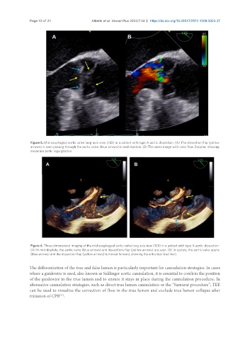
Figure 5. Mid-esophageal aortic valve long-axis view (143) in a patient with type A aortic dissection. (A) The dissection flap (yellow
arrows) is seen passing through the aortic valve (blue arrows) in end-diastole. (B) The same image with color flow Doppler, showing
moderate aortic regurgitation.
Figure 6. Three-dimensional imaging of the mid-esophageal aortic valve long axis view (128) in a patient with type A aortic dissection.
(A) In mid-diastole, the aortic valve (blue arrows) and dissections flap (yellow arrows) are seen. (B) In systole, the aortic valve opens
(blue arrows) and the dissection flap (yellow arrows) is moved forward, showing the entry tear (red line).
The differentiation of the true and false lumen is particularly important for cannulation strategies. In cases
where a guidewire is used, also known as Seldinger aortic cannulation, it is essential to confirm the position
of the guidewire in the true lumen and to ensure it stays in place during the cannulation procedure. In
alternative cannulation strategies, such as direct true lumen cannulation or the “Samurai procedure”, TEE
can be used to visualize the correction of flow in the true lumen and exclude true lumen collapse after
[71]
initiation of CPB .