
Page 166 - Read Online
P. 166
Alberts et al. Vessel Plus 2023;7:34 https://dx.doi.org/10.20517/2574-1209.2023.37 Page 5 of 24
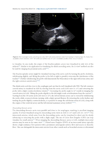
Figure 2. Mid-esophageal aortic valve long axis view (127) with measurements made from leading-to-leading edge. Ao: Aorta; Asc:
ascendens; Diam: diameter; LVOT: left ventricular outflow tract; ST junct: sinotubular junction; SVals: sinus of valsalva.
to visualize. In one study, the origin of the brachiocephalic artery was visualized in only 36% of the
subjects . Similar to its application in visualizing the distal ascending aorta, the A-view® method can also
[38]
be used for imaging the proximal aortic arch [35,36] .
The brachiocephalic artery might be visualized starting at the aortic arch by turning the probe clockwise,
withdrawing slightly, and tilting the probe to the left or right to possibly overcome the interference of the
trachea . Further withdrawing the probe will demonstrate the bifurcation to the right subclavian artery and
[39]
the right common carotid artery [38,39] .
The distal aortic arch lies close to the esophagus and can thus be well visualized with TEE. The left common
carotid artery is visualized in SAX by starting from the aortic arch LAX view (0-10°) and retracting the
[31]
probe with a slight counterclockwise rotation . Increasing the probe angle to 90° results in imaging the
carotid artery in LAX. Tilting the probe slightly to the left might evade reverberations from the trachea .
[38]
Starting from the UE aortic arch LAX view (0-10°), the left subclavian artery can be viewed by withdrawing
the probe while counterclockwise turning, with a slight upward flexion. By retracting the probe further and
turning the probe slightly counterclockwise, it is possible to image the subclavian artery in LAX, along with
the origins of the vertebral artery and the left internal mammary artery (LIMA) [31,38,40] .
Descending thoracic aorta
The descending thoracic aorta runs parallel and close to the esophagus, resulting in excellent imaging
quality. It is best visualized using the descending aorta views in SAX (0-10°) and LAX (90-100°) . The
[12]
intercostal arteries, which arise from the descending aorta, can be visualized in short axis by slowly
advancing or retracting the probe with a slight angle. The use of Color flow Doppler (CFD) can help
visualize the small arteries . When imaging the descending thoracic aorta in LAX, multiple intercostal
[41]
arteries may be seen in the same view [12,31] . Pulsed wave Doppler (PWD) of an intercostal artery typically
shows a high resistance flow pattern with a prominent anterograde systolic flow and nearly no diastolic
flow. This can distinguish the intercostal arteries from other vessels such as bronchial arteries or abdominal